


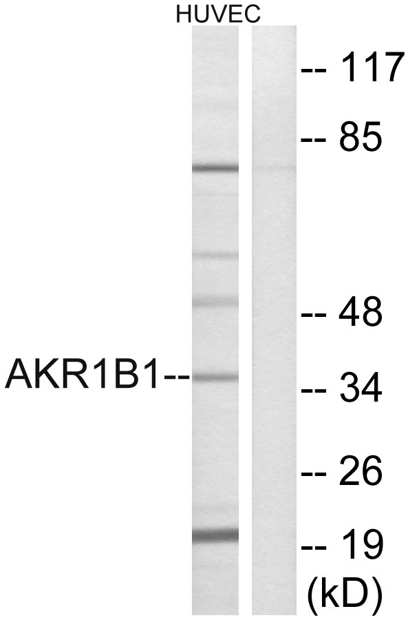
Figure 1. Western blot analysis of AKR1B1 using anti-AKR1B1 antibody (PB10035). Electrophoresis was performed on a 5-20% SDS-PAGE gel at 70V (Stacking gel) / 90V (Resolving gel) for 2-3 hours. The sample well of each lane was loaded with 30 ug of sample under reducing conditions. Lane 1: human SiHa whole cell lysates, Lane 2: rat testis tissue lysates, Lane 3: mouse NIH/3T3 whole cell lysates. After electrophoresis, proteins were transferred to a nitrocellulose membrane at 150 mA for 50-90 minutes. Blocked the membrane with 5% non-fat milk/TBS for 1.5 hour at RT. The membrane was incubated with rabbit anti-AKR1B1 antigen affinity purified polyclonal antibody (Catalog # PB10035) at 0.5 microg/mL overnight at 4°C, then washed with TBS-0.1%Tween 3 times with 5 minutes each and probed with a goat anti-rabbit IgG-HRP secondary antibody at a dilution of 1:5000 for 1.5 hour at RT. The signal is developed using an Enhanced Chemiluminescent detection (ECL) kit (Catalog # EK1002) with Tanon 5200 system. A specific band was detected for AKR1B1 at approximately 36 kDa. The expected band size for AKR1B1 is at 36 kDa.
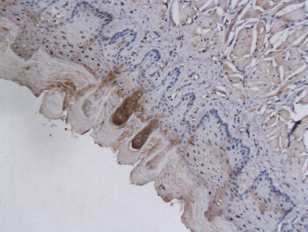
. AKR1B1 was detected in paraffin-embedded section of rat intestine tissues. Heat mediated antigen retrieval was performed in citrate buffer (pH6, epitope retrieval solution) for 20 mins. The tissue section was blocked with 10% goat serum. The tissue section was then incubated with 1microg/ml rabbit anti-AKR1B1 Antibody (PB10035) overnight at 4°C. Biotinylated goat anti-rabbit IgG was used as secondary antibody and incubated for 30 minutes at 37°C. The tissue section was developed using Strepavidin-Biotin-Complex (SABC)(Catalog # SA1022) with DAB as the chromogen.")
. AKR1B1 was detected in paraffin-embedded section of rat adrenal gland tissues. Heat mediated antigen retrieval was performed in citrate buffer (pH6, epitope retrieval solution) for 20 mins. The tissue section was blocked with 10% goat serum. The tissue section was then incubated with 1microg/ml rabbit anti-AKR1B1 Antibody (PB10035) overnight at 4°C. Biotinylated goat anti-rabbit IgG was used as secondary antibody and incubated for 30 minutes at 37°C. The tissue section was developed using Strepavidin-Biotin-Complex (SABC)(Catalog # SA1022) with DAB as the chromogen.")
. AKR1B1 was detected in paraffin-embedded section of human placenta tissues. Heat mediated antigen retrieval was performed in citrate buffer (pH6, epitope retrieval solution) for 20 mins. The tissue section was blocked with 10% goat serum. The tissue section was then incubated with 1microg/ml rabbit anti-AKR1B1 Antibody (PB10035) overnight at 4°C. Biotinylated goat anti-rabbit IgG was used as secondary antibody and incubated for 30 minutes at 37°C. The tissue section was developed using Strepavidin-Biotin-Complex (SABC)(Catalog # SA1022) with DAB as the chromogen.")
. Overlay histogram showing U20S cells stained with PB10035 (Blue line). To facilitate intracellular staining, cells were fixed with 4% paraformaldehyde and permeabilized with permeabilization buffer. The cells were blocked with 10% normal goat serum. And then incubated with rabbit anti-AKR1B1 Antibody (PB10035,1microg/1x106 cells) for 30 min at 20°C. DyLight®488 conjugated goat anti-rabbit IgG (BA1127, 5-10microg/1x106 cells) was used as secondary antibody for 30 minutes at 20°C. Isotype control antibody (Green line) was rabbit IgG (1microg/1x106) used under the same conditions. Unlabelled sample without incubation with primary antibody and secondary antibody (Red line) was used as a blank control.")
Figure 1. Western blot analysis of AKR1B1 using anti-AKR1B1 antibody (PB10035). Electrophoresis was performed on a 5-20% SDS-PAGE gel at 70V (Stacking gel) / 90V (Resolving gel) for 2-3 hours. The sample well of each lane was loaded with 30 ug of sample under reducing conditions. Lane 1: human SiHa whole cell lysates, Lane 2: rat testis tissue lysates, Lane 3: mouse NIH/3T3 whole cell lysates. After electrophoresis, proteins were transferred to a nitrocellulose membrane at 150 mA for 50-90 minutes. Blocked the membrane with 5% non-fat milk/TBS for 1.5 hour at RT. The membrane was incubated with rabbit anti-AKR1B1 antigen affinity purified polyclonal antibody (Catalog # PB10035) at 0.5 microg/mL overnight at 4°C, then washed with TBS-0.1%Tween 3 times with 5 minutes each and probed with a goat anti-rabbit IgG-HRP secondary antibody at a dilution of 1:5000 for 1.5 hour at RT. The signal is developed using an Enhanced Chemiluminescent detection (ECL) kit (Catalog # EK1002) with Tanon 5200 system. A specific band was detected for AKR1B1 at approximately 36 kDa. The expected band size for AKR1B1 is at 36 kDa.
Anti-AKR1B1 Antibody Picoband(r)
PB10035-CARRIER-FREE
ApplicationsFlow Cytometry, Western Blot, ImmunoHistoChemistry
Product group Antibodies
ReactivityHuman, Mouse, Rat
TargetAKR1B1
Overview
- SupplierBoster Bio
- Product NameAnti-AKR1B1 Antibody Picoband(r)
- Delivery Days Customer9
- Application Supplier NoteTested Species: In-house tested species with positive results. By Heat: Boiling the paraffin sections in 10mM citrate buffer, pH6.0, for 20mins is required for the staining of formalin/paraffin sections. Other applications have not been tested. Optimal dilutions should be determined by end users.
- ApplicationsFlow Cytometry, Western Blot, ImmunoHistoChemistry
- CertificationResearch Use Only
- ClonalityPolyclonal
- Concentration500 ug/ml
- Gene ID231
- Target nameAKR1B1
- Target descriptionaldo-keto reductase family 1 member B
- Target synonymsADR, ALDR1, ALR2, AR, aldo-keto reductase family 1 member B1, Lii5-2 CTCL tumor antigen, aldehyde reductase 1, aldose reductase, low Km aldose reductase
- HostRabbit
- IsotypeIgG
- Protein IDP15121
- Protein NameAldo-keto reductase family 1 member B1
- Scientific DescriptionBoster Bio Anti-AKR1B1 Antibody Picoband® catalog # PB10035. Tested in Flow Cytometry, IHC, WB applications. This antibody reacts with Human, Mouse, Rat. The brand Picoband indicates this is a premium antibody that guarantees superior quality, high affinity, and strong signals with minimal background in Western blot applications. Only our best-performing antibodies are designated as Picoband, ensuring unmatched performance.
- ReactivityHuman, Mouse, Rat
- Storage Instruction-20°C,2°C to 8°C
- UNSPSC12352203
Related products
Product group Antibodies
Anti-AKR1B1 AntibodyA99317
ApplicationsWestern Blot, ELISA
ReactivityHuman, Rat
- SizePrice
Product group Antibodies
Anti-AKR1B1 Antibody144-60529
ApplicationsWestern Blot, ImmunoHistoChemistry
ReactivityHuman, Mouse, Rat
TargetAKR1B1
- SizePrice
Product group Antibodies
AKR1B1 Polyclonal AntibodyBS-2405R
ApplicationsImmunoFluorescence, Western Blot, ELISA, ImmunoCytoChemistry, ImmunoHistoChemistry, ImmunoHistoChemistry Frozen, ImmunoHistoChemistry Paraffin
ReactivityBovine, Canine, Human, Mouse, Rabbit, Rat
TargetAKR1B1
- SizePrice
Product group Antibodies
AKR1B1 AntibodyCSB-PA001539LA01HU
ApplicationsImmunoFluorescence, Western Blot, ELISA, ImmunoHistoChemistry
ReactivityHuman
TargetAKR1B1
- SizePrice
Product group Antibodies
AKR1B1 Polyclonal AntibodyCAC14579
ApplicationsImmunoFluorescence, Western Blot, ELISA, ImmunoHistoChemistry
TargetAKR1B1
- SizePrice

Product group Antibodies
Anti-AKR1B1 AntibodyHPA026425
ApplicationsImmunoCytoChemistry, ImmunoHistoChemistry
ReactivityHuman
TargetAKR1B1
- SizePrice
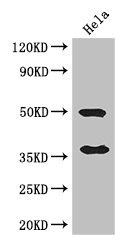
![Various whole cell extracts (30 μg) were separated by 10% SDS-PAGE, and the membrane was blotted with AKR1B1 antibody [N1C3] (GTX113381) diluted at 1:1000. The HRP-conjugated anti-rabbit IgG antibody (GTX213110-01) was used to detect the primary antibody. Corresponding RNA expression data for the same cell lines are based on Human Protein Atlas program.](https://www.genetex.com/upload/website/prouct_img/normal/GTX113381/GTX113381_43936_20200501_WB_TPM_watermark_w_23060501_162.webp)
Product group Antibodies
AKR1B1 antibody [N1C3]GTX113381
ApplicationsImmunoFluorescence, Western Blot, ImmunoCytoChemistry, ImmunoHistoChemistry, ImmunoHistoChemistry Paraffin
ReactivityHuman, Porcine, Rat
TargetAKR1B1
- SizePrice
Product group Antibodies

AKR1B1 / Aldose Reductase AntibodyLS-C400454
ApplicationsWestern Blot, ELISA, ImmunoHistoChemistry
ReactivityHuman, Mouse, Rat
TargetAKR1B1
- SizePrice
Product group Antibodies
Anti-AKR1B1 AntibodyCAB13944
ApplicationsWestern Blot, ELISA
ReactivityHuman
TargetAKR1B1
- SizePrice