


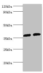
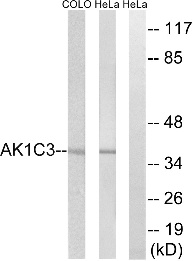
Figure 1. Western blot analysis of AKR1C3 using anti-AKR1C3 antibody (A01820-1). Electrophoresis was performed on a 5-20% SDS-PAGE gel at 70V (Stacking gel) / 90V (Resolving gel) for 2-3 hours. The sample well of each lane was loaded with 30 ug of sample under reducing conditions. Lane 1: human HCCP tissue lysates, Lane 2: human A549 whole cell lysates. After electrophoresis, proteins were transferred to a nitrocellulose membrane at 150 mA for 50-90 minutes. Blocked the membrane with 5% non-fat milk/TBS for 1.5 hour at RT. The membrane was incubated with rabbit anti-AKR1C3 antigen affinity purified polyclonal antibody (Catalog # A01820-1) at 0.5 microg/mL overnight at 4°C, then washed with TBS-0.1%Tween 3 times with 5 minutes each and probed with a goat anti-rabbit IgG-HRP secondary antibody at a dilution of 1:5000 for 1.5 hour at RT. The signal is developed using an Enhanced Chemiluminescent detection (ECL) kit (Catalog # EK1002) with Tanon 5200 system. A specific band was detected for AKR1C3 at approximately 37 kDa. The expected band size for AKR1C3 is at 37 kDa.
.")
Figure 1. Western blot analysis of AKR1C3 using anti-AKR1C3 antibody (A01820-1). Electrophoresis was performed on a 5-20% SDS-PAGE gel at 70V (Stacking gel) / 90V (Resolving gel) for 2-3 hours. The sample well of each lane was loaded with 30 ug of sample under reducing conditions. Lane 1: human HCCP tissue lysates, Lane 2: human A549 whole cell lysates. After electrophoresis, proteins were transferred to a nitrocellulose membrane at 150 mA for 50-90 minutes. Blocked the membrane with 5% non-fat milk/TBS for 1.5 hour at RT. The membrane was incubated with rabbit anti-AKR1C3 antigen affinity purified polyclonal antibody (Catalog # A01820-1) at 0.5 microg/mL overnight at 4°C, then washed with TBS-0.1%Tween 3 times with 5 minutes each and probed with a goat anti-rabbit IgG-HRP secondary antibody at a dilution of 1:5000 for 1.5 hour at RT. The signal is developed using an Enhanced Chemiluminescent detection (ECL) kit (Catalog # EK1002) with Tanon 5200 system. A specific band was detected for AKR1C3 at approximately 37 kDa. The expected band size for AKR1C3 is at 37 kDa.
Anti-AKR1C3 Antibody Picoband(r)
A01820-1-CARRIER-FREE
ApplicationsFlow Cytometry, Western Blot
Product group Antibodies
ReactivityHuman
TargetAKR1C3
Overview
- SupplierBoster Bio
- Product NameAnti-AKR1C3 Antibody Picoband(r)
- Delivery Days Customer9
- ApplicationsFlow Cytometry, Western Blot
- CertificationResearch Use Only
- ClonalityPolyclonal
- Concentration500 ug/ml
- Gene ID8644
- Target nameAKR1C3
- Target descriptionaldo-keto reductase family 1 member C3
- Target synonymsDD3, DDX, HA1753, HAKRB, HAKRe, HSD17B5, PGFS, hluPGFS, aldo-keto reductase family 1 member C3, 3-alpha hydroxysteroid dehydrogenase, type II, 3-alpha-HSD type II, brain, chlordecone reductase homolog HAKRb, dihydrodiol dehydrogenase 3, dihydrodiol dehydrogenase X, indanol dehydrogenase, prostaglandin F synthase, testosterone 17-beta-dehydrogenase 5, trans-1,2-dihydrobenzene-1,2-diol dehydrogenase, type IIb 3-alpha hydroxysteroid dehydrogenase
- HostRabbit
- IsotypeIgG
- Protein IDP42330
- Protein NameAldo-keto reductase family 1 member C3
- Scientific DescriptionBoster Bio Anti-AKR1C3 Antibody Picoband® catalog # A01820-1. Tested in Flow Cytometry, WB applications. This antibody reacts with Human. The brand Picoband indicates this is a premium antibody that guarantees superior quality, high affinity, and strong signals with minimal background in Western blot applications. Only our best-performing antibodies are designated as Picoband, ensuring unmatched performance.
- ReactivityHuman
- Storage Instruction-20°C,2°C to 8°C
- UNSPSC12352203
Related products
Product group Antibodies
AKR1C3 AntibodyCSB-PA001544ESR1HU
ApplicationsImmunoPrecipitation, Western Blot, ELISA, ImmunoHistoChemistry
ReactivityHuman
TargetAKR1C3
- SizePrice
Product group Antibodies
Anti-AKR1C3 AntibodyA100616
ApplicationsWestern Blot, ELISA
ReactivityHuman
- SizePrice
Product group Antibodies

DDX / AKR1C3 AntibodyLS-C748613
ApplicationsImmunoFluorescence, Western Blot, ImmunoHistoChemistry
ReactivityHuman, Rat
TargetAKR1C3
- SizePrice
Product group Antibodies

Goat anti-AKR1C3EB06637
ApplicationsWestern Blot, ELISA, ImmunoHistoChemistry
ReactivityHuman
TargetAKR1C3
- SizePrice
Product group Antibodies
Akr1C3 Recombinant AntibodyCAC11975
ApplicationsFlow Cytometry, ImmunoFluorescence, ELISA
TargetAKR1C3
- SizePrice
Product group Antibodies
AKR1C3 antibody [C2C3], C-termGTX104627
ApplicationsImmunoFluorescence, ImmunoPrecipitation, Western Blot, ImmunoCytoChemistry, ImmunoHistoChemistry, ImmunoHistoChemistry Paraffin
ReactivityHuman, Rat
TargetAKR1C3
- SizePrice
Product group Antibodies
AKR1C3 Recombinant Antibody, AbBy Fluor-405 ConjugatedBSM-62035R-BF405
ApplicationsFlow Cytometry, Western Blot
ReactivityHuman
TargetAKR1C3
- SizePrice
Product group Antibodies
Anti-AKR1C3 Antibody144-01781
ApplicationsImmunoPrecipitation, Western Blot
ReactivityHuman, Mouse, Rat
TargetAKR1C3
- SizePrice