


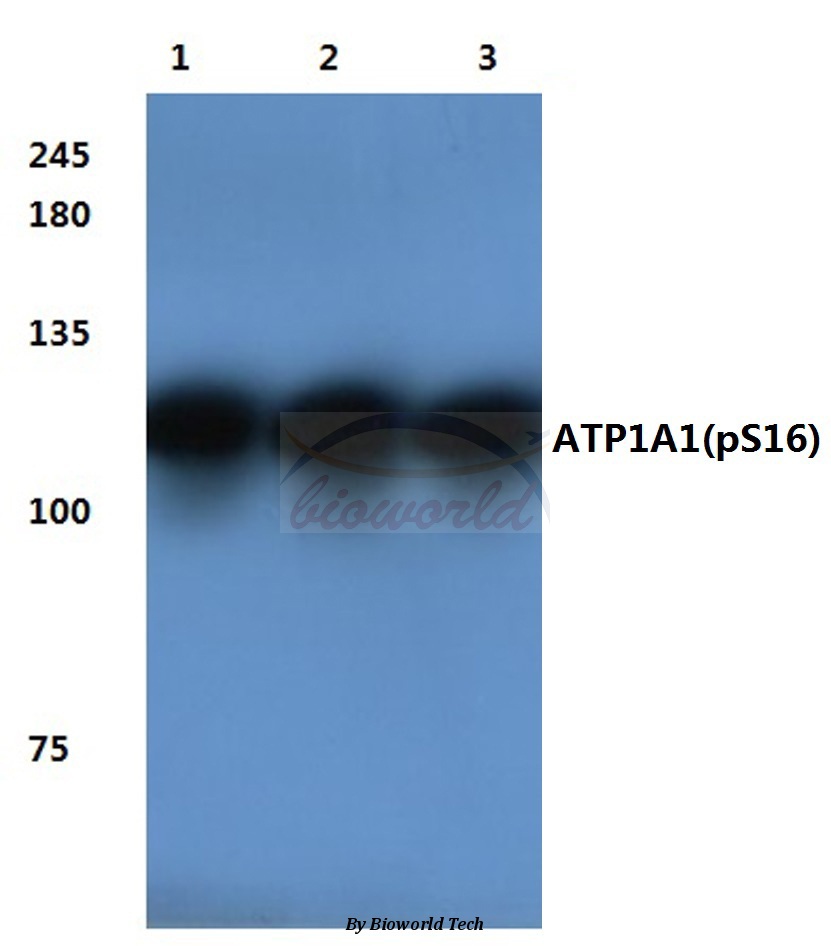
Figure 1. Western blot analysis of ATP1A1 using anti-ATP1A1 antibody (PB9482). Electrophoresis was performed on a 5-20% SDS-PAGE gel at 70V (Stacking gel) / 90V (Resolving gel) for 2-3 hours. The sample well of each lane was loaded with 30 ug of sample under reducing conditions. Lane 1: rat brain tissue lysates, Lane 2: rat brain tissue lysates, Lane 3: mouse brain tissue lysates, Lane 4: mouse brain tissue lysates. After Electrophoresis, proteins were transferred to a Nitrocellulose membrane at 150mA for 50-90 minutes. Blocked the membrane with 5% Non-fat Milk/ TBS for 1.5 hour at RT. The membrane was incubated with rabbit anti-ATP1A1 antigen affinity purified polyclonal antibody (Catalog # PB9482) at 0.5 microg/mL overnight at 4°C, then washed with TBS-0.1%Tween 3 times with 5 minutes each and probed with a goat anti-rabbit IgG-DyLight 647 Conjugated secondary antibody (Left) at a dilution of 1:2000 or a goat anti-rabbit IgG-HRP Conjugated secondary antibody (Right) at a dilution of 1:5000 for 1.5 hour at RT. The signal is developed using an Enhanced Chemiluminescent detection (ECL) kit (Catalog # EK1002) with Tanon 5200 system. A specific band was detected for ATP1A1 approximately 100 kDa. The expected band size for ATP1A1 is at 74, 110, 113 kDa.
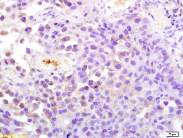
. ATP1A1 was detected in a paraffin-embedded section of human ovarian cancer tissue. Heat mediated antigen retrieval was performed in EDTA buffer (pH 8.0, epitope retrieval solution). The tissue section was blocked with 10% goat serum. The tissue section was then incubated with 2 microg/ml rabbit anti-ATP1A1 Antibody (PB9482) overnight at 4°C. Peroxidase Conjugated Goat Anti-rabbit IgG was used as secondary antibody and incubated for 30 minutes at 37°C. The tissue section was developed using HRP Conjugated Rabbit IgG Super Vision Assay Kit (Catalog # SV0002) with DAB as the chromogen.")
. ATP1A1 was detected in a paraffin-embedded section of human colon cancer tissue. Heat mediated antigen retrieval was performed in EDTA buffer (pH 8.0, epitope retrieval solution). The tissue section was blocked with 10% goat serum. The tissue section was then incubated with 2 microg/ml rabbit anti-ATP1A1 Antibody (PB9482) overnight at 4°C. Peroxidase Conjugated Goat Anti-rabbit IgG was used as secondary antibody and incubated for 30 minutes at 37°C. The tissue section was developed using HRP Conjugated Rabbit IgG Super Vision Assay Kit (Catalog # SV0002) with DAB as the chromogen.")
. ATP1A1 was detected in a paraffin-embedded section of human endometrioid adenocarcinoma tissue. Heat mediated antigen retrieval was performed in EDTA buffer (pH 8.0, epitope retrieval solution). The tissue section was blocked with 10% goat serum. The tissue section was then incubated with 2 microg/ml rabbit anti-ATP1A1 Antibody (PB9482) overnight at 4°C. Peroxidase Conjugated Goat Anti-rabbit IgG was used as secondary antibody and incubated for 30 minutes at 37°C. The tissue section was developed using HRP Conjugated Rabbit IgG Super Vision Assay Kit (Catalog # SV0002) with DAB as the chromogen.")
. ATP1A1 was detected in a paraffin-embedded section of human liver cancer tissue. Heat mediated antigen retrieval was performed in EDTA buffer (pH 8.0, epitope retrieval solution). The tissue section was blocked with 10% goat serum. The tissue section was then incubated with 2 microg/ml rabbit anti-ATP1A1 Antibody (PB9482) overnight at 4°C. Peroxidase Conjugated Goat Anti-rabbit IgG was used as secondary antibody and incubated for 30 minutes at 37°C. The tissue section was developed using HRP Conjugated Rabbit IgG Super Vision Assay Kit (Catalog # SV0002) with DAB as the chromogen.")
. ATP1A1 was detected in a paraffin-embedded section of human spleen tissue. Heat mediated antigen retrieval was performed in EDTA buffer (pH 8.0, epitope retrieval solution). The tissue section was blocked with 10% goat serum. The tissue section was then incubated with 2 microg/ml rabbit anti-ATP1A1 Antibody (PB9482) overnight at 4°C. Peroxidase Conjugated Goat Anti-rabbit IgG was used as secondary antibody and incubated for 30 minutes at 37°C. The tissue section was developed using HRP Conjugated Rabbit IgG Super Vision Assay Kit (Catalog # SV0002) with DAB as the chromogen.")
. ATP1A1 was detected in a paraffin-embedded section of mouse kidney tissue. Heat mediated antigen retrieval was performed in EDTA buffer (pH 8.0, epitope retrieval solution). The tissue section was blocked with 10% goat serum. The tissue section was then incubated with 2 microg/ml rabbit anti-ATP1A1 Antibody (PB9482) overnight at 4°C. Peroxidase Conjugated Goat Anti-rabbit IgG was used as secondary antibody and incubated for 30 minutes at 37°C. The tissue section was developed using HRP Conjugated Rabbit IgG Super Vision Assay Kit (Catalog # SV0002) with DAB as the chromogen.")
. ATP1A1 was detected in a paraffin-embedded section of mouse kidney tissue. Heat mediated antigen retrieval was performed in EDTA buffer (pH 8.0, epitope retrieval solution). The tissue section was blocked with 10% goat serum. The tissue section was then incubated with 2 microg/ml rabbit anti-ATP1A1 Antibody (PB9482) overnight at 4°C. Peroxidase Conjugated Goat Anti-rabbit IgG was used as secondary antibody and incubated for 30 minutes at 37°C. The tissue section was developed using HRP Conjugated Rabbit IgG Super Vision Assay Kit (Catalog # SV0002) with DAB as the chromogen.")
. ATP1A1 was detected in a paraffin-embedded section of rat kidney tissue. Heat mediated antigen retrieval was performed in EDTA buffer (pH 8.0, epitope retrieval solution). The tissue section was blocked with 10% goat serum. The tissue section was then incubated with 2 microg/ml rabbit anti-ATP1A1 Antibody (PB9482) overnight at 4°C. Peroxidase Conjugated Goat Anti-rabbit IgG was used as secondary antibody and incubated for 30 minutes at 37°C. The tissue section was developed using HRP Conjugated Rabbit IgG Super Vision Assay Kit (Catalog # SV0002) with DAB as the chromogen.")
Figure 1. Western blot analysis of ATP1A1 using anti-ATP1A1 antibody (PB9482). Electrophoresis was performed on a 5-20% SDS-PAGE gel at 70V (Stacking gel) / 90V (Resolving gel) for 2-3 hours. The sample well of each lane was loaded with 30 ug of sample under reducing conditions. Lane 1: rat brain tissue lysates, Lane 2: rat brain tissue lysates, Lane 3: mouse brain tissue lysates, Lane 4: mouse brain tissue lysates. After Electrophoresis, proteins were transferred to a Nitrocellulose membrane at 150mA for 50-90 minutes. Blocked the membrane with 5% Non-fat Milk/ TBS for 1.5 hour at RT. The membrane was incubated with rabbit anti-ATP1A1 antigen affinity purified polyclonal antibody (Catalog # PB9482) at 0.5 microg/mL overnight at 4°C, then washed with TBS-0.1%Tween 3 times with 5 minutes each and probed with a goat anti-rabbit IgG-DyLight 647 Conjugated secondary antibody (Left) at a dilution of 1:2000 or a goat anti-rabbit IgG-HRP Conjugated secondary antibody (Right) at a dilution of 1:5000 for 1.5 hour at RT. The signal is developed using an Enhanced Chemiluminescent detection (ECL) kit (Catalog # EK1002) with Tanon 5200 system. A specific band was detected for ATP1A1 approximately 100 kDa. The expected band size for ATP1A1 is at 74, 110, 113 kDa.
Anti-alpha 1 Sodium Potassium ATPase/ATP1A1 Antibody Picoband(r)
PB9482-CARRIER-FREE
ApplicationsImmunoFluorescence, Western Blot, ImmunoHistoChemistry
Product group Antibodies
ReactivityHuman, Mouse, Rat
TargetATP1A1
Overview
- SupplierBoster Bio
- Product NameAnti-alpha 1 Sodium Potassium ATPase/ATP1A1 Antibody Picoband(r)
- Delivery Days Customer9
- Application Supplier NoteTested Species: In-house tested species with positive results. Other applications have not been tested. Optimal dilutions should be determined by end users.
- ApplicationsImmunoFluorescence, Western Blot, ImmunoHistoChemistry
- CertificationResearch Use Only
- ClonalityPolyclonal
- Concentration500 ug/ml
- Gene ID476
- Target nameATP1A1
- Target descriptionATPase Na+/K+ transporting subunit alpha 1
- Target synonymsCMT2DD, HOMGSMR2, sodium/potassium-transporting ATPase subunit alpha-1, ATPase, Na+/K+ transporting, alpha 1 polypeptide, Na(+)/K(+) ATPase alpha-1 subunit, Na+/K+ ATPase 1, Na, K-ATPase, alpha-A catalytic polypeptide, Na,K-ATPase alpha-1 subunit, Na,K-ATPase catalytic subunit alpha-A protein, sodium pump subunit alpha-1, sodium-potassium ATPase catalytic subunit alpha-1, sodium-potassium-ATPase, alpha 1 polypeptide
- HostRabbit
- IsotypeIgG
- Protein IDP05023
- Protein NameSodium/potassium-transporting ATPase subunit alpha-1
- Scientific DescriptionBoster Bio Anti-alpha 1 Sodium Potassium ATPase/ATP1A1 Antibody Picoband® catalog # PB9482. Tested in IF, IHC, WB applications. This antibody reacts with Human, Mouse, Rat. The brand Picoband indicates this is a premium antibody that guarantees superior quality, high affinity, and strong signals with minimal background in Western blot applications. Only our best-performing antibodies are designated as Picoband, ensuring unmatched performance.
- ReactivityHuman, Mouse, Rat
- Storage Instruction-20°C,2°C to 8°C
- UNSPSC12352203
Related products
Product group Antibodies
ApplicationsWestern Blot, ImmunoHistoChemistry
ReactivityHuman, Mouse, Rat
- SizePrice
Product group Antibodies

ATP1A1 Antibody (Tyr260)LS-C769387
ApplicationsWestern Blot, ELISA
ReactivityHuman, Mouse, Rat
TargetATP1A1
- SizePrice
Product group Antibodies
References
ATP1A1 Polyclonal Antibodybs-4255R
ApplicationsWestern Blot, ELISA, ImmunoHistoChemistry, ImmunoHistoChemistry Paraffin
ReactivityChicken, Guinea Pig, Human, Mouse, Porcine, Rabbit, Rat
TargetATP1A1
- SizePrice
Product group Antibodies
ATP1A1 AntibodyCSB-PA003364
ApplicationsWestern Blot, ELISA
ReactivityHuman, Mouse, Rat
TargetATP1A1
- SizePrice
Product group Antibodies
ApplicationsImmunoPrecipitation, Western Blot, ImmunoCytoChemistry, ImmunoHistoChemistry
ReactivityMouse, Porcine, Rat
TargetATP1A1
- SizePrice
![ICC/IF analysis of HeLa cells using GTX22872 Sodium/Potassium ATPase alpha 1 antibody [M8-P1-A3]. Green : Primary antibody Blue : Nuclei Red : Actin Fixation : Formalin Permeabilization : 0.1% Triton X-100 in TBS for 15 minutes at room temperature Dilution : 1:100 for at least 1 hour at room temperature](https://www.genetex.com/upload/website/prouct_img/normal/GTX22872/GTX22872_532_ICC-IF_w_23060620_863.webp)
Product group Antibodies
References
ApplicationsFlow Cytometry, ImmunoFluorescence, Western Blot, ImmunoCytoChemistry, ImmunoHistoChemistry, ImmunoHistoChemistry Frozen, ImmunoHistoChemistry Paraffin, Neutralisation/Blocking
ReactivityCanine, Human, Mouse, Porcine, Primate, Rat, Sheep, Yeast
TargetATP1A1
- SizePrice