


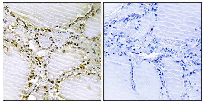
Immunohistochemical staining of human skeletal muscle shows strong cytoplasmic positivity.
Immunohistochemical staining of human skeletal muscle shows strong cytoplasmic positivity.
Anti-AMPD1 Antibody
HPA026478
ApplicationsImmunoHistoChemistry
Product group Antibodies
ReactivityHuman
TargetAMPD1
Overview
- SupplierAtlas Antibodies
- Product NameAnti-AMPD1 Antibody
- Delivery Days Customer4
- ApplicationsImmunoHistoChemistry
- CertificationResearch Use Only
- ClonalityPolyclonal
- ConjugateUnconjugated
- Gene ID270
- Target nameAMPD1
- Target descriptionadenosine monophosphate deaminase 1
- Target synonymsMAD, MADA, MMDD, AMP deaminase 1, AMPD, adenosine monophosphate deaminase 1 (isoform M), adenosine monophosphate deaminase-1 (muscle), myoadenylate deaminase, skeletal muscle AMPD
- HostRabbit
- IsotypeIgG
- Protein IDP23109
- Protein NameAMP deaminase 1
- Scientific DescriptionRecombinant Protein Epitope Signature Tag (PrEST) antigen sequence
- ReactivityHuman
- Storage Instruction-20°C,2°C to 8°C
- UNSPSC41116161
Datasheet
MSDS
Related products
Product group Antibodies
Anti-AMPD1 AntibodyA97051
ApplicationsELISA, ImmunoHistoChemistry
ReactivityHuman, Mouse, Rat
- SizePrice

Product group Antibodies
Anti-AMPD1 Antibody Picoband(r)A04209-1-CARRIER-FREE
ApplicationsFlow Cytometry, Western Blot, ELISA, ImmunoHistoChemistry
ReactivityHuman, Mouse, Rat
TargetAMPD1
- SizePrice
Product group Antibodies
Anti-AMPD1 Antibody144-03584
ApplicationsWestern Blot
ReactivityHuman, Mouse, Rat
TargetAMPD1
- SizePrice
Product group Antibodies
AMPD1 AntibodyCSB-PA001680LA01HU
ApplicationsWestern Blot, ELISA
ReactivityHuman
TargetAMPD1
- SizePrice
Product group Antibodies

Goat anti-AMPD1EB08258
ApplicationsELISA
ReactivityBovine, Canine, Human, Mouse, Porcine, Rat
TargetAMPD1
- SizePrice
Product group Antibodies
AMPD1 Polyclonal AntibodyCAC15036
ApplicationsWestern Blot, ELISA
TargetAMPD1
- SizePrice
Product group Antibodies

AMPD1 AntibodyLS-C402884
ApplicationsWestern Blot, ELISA, ImmunoHistoChemistry
ReactivityHuman
TargetAMPD1
- SizePrice

Product group Antibodies
Anti-AMPD1 AntibodyHPA028080
ApplicationsWestern Blot, ImmunoHistoChemistry
ReactivityHuman
TargetAMPD1
- SizePrice