


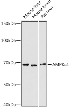
Figure 1. Western blot analysis of AMPK alpha 1/PRKAA1 using anti-AMPK alpha 1/PRKAA1 antibody (A00994-6). Electrophoresis was performed on a 5-20% SDS-PAGE gel at 70V (Stacking gel) / 90V (Resolving gel) for 2-3 hours. The sample well of each lane was loaded with 50ug of sample under reducing conditions. Lane 1: human Jurkat whole cell lysates, Lane 2: human Hela whole cell lysates, Lane 3: human MCF-7 whole cell lysates, Lane 4: monkey Cos-7 whole cell lysates, Lane 5: human K572 whole cell lysates, Lane 6: human HEK293 whole cell lysates, Lane 7: rat brain tissue lysates, Lane 8: rat heart tissue lysates, Lane 9: mouse brain tissue lysates, Lane 10: mouse heart tissue lysates, Lane 11: mouse intestines tissue lysates. After Electrophoresis, proteins were transferred to a Nitrocellulose membrane at 150mA for 50-90 minutes. Blocked the membrane with 5% Non-fat Milk/ TBS for 1.5 hour at RT. The membrane was incubated with rabbit anti-AMPK alpha 1/PRKAA1 antigen affinity purified polyclonal antibody (Catalog # A00994-6) at 0.25 microg/mL overnight at 4°C, then washed with TBS-0.1%Tween 3 times with 5 minutes each and probed with a goat anti-rabbit IgG-HRP secondary antibody at a dilution of 1:5000 for 1.5 hour at RT. The signal is developed using an Enhanced Chemiluminescent detection (ECL) kit (Catalog # EK1002) with Tanon 5200 system. A specific band was detected for AMPK alpha 1/PRKAA1 at approximately 64-66KD. The expected band size for AMPK alpha 1/PRKAA1 is at 64KD.
. Overlay histogram showing SiHa cells stained with A00994-6 (Blue line). To facilitate intracellular staining, cells were fixed with 4% paraformaldehyde and permeabilized with permeabilization buffer. The cells were blocked with 10% normal goat serum. And then incubated with rabbit anti-AMPK alpha 1/PRKAA1 Antibody (A00994-6, 1microg/1x106 cells) for 30 min at 20°C. DyLight®488 conjugated goat anti-rabbit IgG (BA1127, 5-10microg/1x106 cells) was used as secondary antibody for 30 minutes at 20°C. Isotype control antibody (Green line) was rabbit IgG (1microg/1x106) used under the same conditions. Unlabelled sample without incubation with primary antibody and secondary antibody (Red line) was used as a blank control.")
Figure 1. Western blot analysis of AMPK alpha 1/PRKAA1 using anti-AMPK alpha 1/PRKAA1 antibody (A00994-6). Electrophoresis was performed on a 5-20% SDS-PAGE gel at 70V (Stacking gel) / 90V (Resolving gel) for 2-3 hours. The sample well of each lane was loaded with 50ug of sample under reducing conditions. Lane 1: human Jurkat whole cell lysates, Lane 2: human Hela whole cell lysates, Lane 3: human MCF-7 whole cell lysates, Lane 4: monkey Cos-7 whole cell lysates, Lane 5: human K572 whole cell lysates, Lane 6: human HEK293 whole cell lysates, Lane 7: rat brain tissue lysates, Lane 8: rat heart tissue lysates, Lane 9: mouse brain tissue lysates, Lane 10: mouse heart tissue lysates, Lane 11: mouse intestines tissue lysates. After Electrophoresis, proteins were transferred to a Nitrocellulose membrane at 150mA for 50-90 minutes. Blocked the membrane with 5% Non-fat Milk/ TBS for 1.5 hour at RT. The membrane was incubated with rabbit anti-AMPK alpha 1/PRKAA1 antigen affinity purified polyclonal antibody (Catalog # A00994-6) at 0.25 microg/mL overnight at 4°C, then washed with TBS-0.1%Tween 3 times with 5 minutes each and probed with a goat anti-rabbit IgG-HRP secondary antibody at a dilution of 1:5000 for 1.5 hour at RT. The signal is developed using an Enhanced Chemiluminescent detection (ECL) kit (Catalog # EK1002) with Tanon 5200 system. A specific band was detected for AMPK alpha 1/PRKAA1 at approximately 64-66KD. The expected band size for AMPK alpha 1/PRKAA1 is at 64KD.
Anti-AMPK alpha 1/PRKAA1 Antibody Picoband(r)
A00994-6-DYLIGHT488
ApplicationsFlow Cytometry, Western Blot, ELISA
Product group Antibodies
ReactivityHuman, Monkey, Mouse, Rat
TargetPRKAA1
Overview
- SupplierBoster Bio
- Product NameAnti-AMPK alpha 1/PRKAA1 Antibody Picoband(r)
- Delivery Days Customer9
- ApplicationsFlow Cytometry, Western Blot, ELISA
- CertificationResearch Use Only
- ClonalityPolyclonal
- Concentration500 ug/ml
- ConjugateDyLight 488
- Gene ID5562
- Target namePRKAA1
- Target descriptionprotein kinase AMP-activated catalytic subunit alpha 1
- Target synonymsAMPK, AMPK alpha 1, AMPKa1, 5'-AMP-activated protein kinase catalytic subunit alpha-1, 5'-AMP-activated protein kinase, catalytic alpha-1 chain, ACACA kinase, AMP -activate kinase alpha 1 subunit, AMP-activated protein kinase, catalytic, alpha-1, AMPK subunit alpha-1, AMPK, alpha, 1, HMGCR kinase, acetyl-CoA carboxylase kinase, hydroxymethylglutaryl-CoA reductase kinase, protein kinase, AMP-activated, alpha 1 catalytic subunit, tau-protein kinase PRKAA1
- HostRabbit
- IsotypeIgG
- Protein IDQ13131
- Protein Name5'-AMP-activated protein kinase catalytic subunit alpha-1
- Scientific DescriptionBoster Bio Anti-AMPK alpha 1/PRKAA1 Antibody Picoband® catalog # A00994-6. Tested in ELISA, Flow Cytometry, WB applications. This antibody reacts with Human, Monkey, Mouse, Rat. The brand Picoband indicates this is a premium antibody that guarantees superior quality, high affinity, and strong signals with minimal background in Western blot applications. Only our best-performing antibodies are designated as Picoband, ensuring unmatched performance.
- ReactivityHuman, Monkey, Mouse, Rat
- Storage Instruction-20°C,2°C to 8°C
- UNSPSC12352203
Related products
Product group Antibodies
PRKAA1 Polyclonal AntibodyCAC14615
ApplicationsImmunoFluorescence, Western Blot, ELISA, ImmunoHistoChemistry
ReactivityMouse
TargetPRKAA1
- SizePrice
Product group Antibodies
References
AMPK alpha-1 Polyclonal AntibodyBS-10344R
ApplicationsFlow Cytometry, Western Blot, ELISA, ImmunoHistoChemistry, ImmunoHistoChemistry Paraffin
ReactivityBovine, Human, Mouse, Rabbit, Rat, Sheep
TargetPRKAA1
- SizePrice
Product group Antibodies

ApplicationsWestern Blot, ELISA
ReactivityHuman
TargetPRKAA1
- SizePrice
Product group Antibodies
ApplicationsImmunoFluorescence, Western Blot, ImmunoCytoChemistry
ReactivityHuman, Mouse, Rat
- SizePrice
Product group Antibodies
anti-AMPK alpha-1 (human), Rabbit Monoclonal (RM301)REV-31-1186-00
ApplicationsWestern Blot, ImmunoHistoChemistry
ReactivityHuman
TargetPRKAA1
- SizePrice

Product group Antibodies
Anti-PRKAA1 AntibodyHPA035409
ApplicationsImmunoCytoChemistry
ReactivityHuman
TargetPRKAA1
- SizePrice
Product group Antibodies
PRKAA1 AntibodyCSB-PA071831
ApplicationsWestern Blot, ELISA, ImmunoHistoChemistry
ReactivityHuman, Mouse, Rat
TargetPRKAA1
- SizePrice