


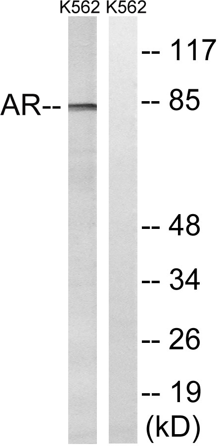
Figure 1. Western blot analysis of Androgen Receptor/AR using anti-Androgen Receptor/AR antibody (A00542). Electrophoresis was performed on a 5-20% SDS-PAGE gel at 70V (Stacking gel) / 90V (Resolving gel) for 2-3 hours. The sample well of each lane was loaded with 50ug of sample under reducing conditions. Lane 1: human K562 whole cell lysates, Lane 2: human U20S whole cell lysates, Lane 3: human HEK293 whole cell lysates, Lane 4: human PC-3 whole cell lysates, Lane 5: human 22RV1 whole cell lysates, Lane 6: human HepG2 whole cell lysates, Lane 7: human CACO-2 whole cell lysates. After Electrophoresis, proteins were transferred to a Nitrocellulose membrane at 150mA for 50-90 minutes. Blocked the membrane with 5% Non-fat Milk/ TBS for 1.5 hour at RT. The membrane was incubated with rabbit anti-Androgen Receptor/AR antigen affinity purified polyclonal antibody (Catalog # A00542) at 0.25 microg/mL overnight at 4°C, then washed with TBS-0.1%Tween 3 times with 5 minutes each and probed with a goat anti-rabbit IgG-HRP secondary antibody at a dilution of 1:5000 for 1.5 hour at RT. The signal is developed using an Enhanced Chemiluminescent detection (ECL) kit (Catalog # EK1002) with Tanon 5200 system. A specific band was detected for Androgen Receptor/AR at approximately 120KD. The expected band size for Androgen Receptor/AR is at 120KD.
. Overlay histogram showing A549 cells stained with A00542 (Blue line).To facilitate intracellular staining, cells were fixed with 4% paraformaldehyde and permeabilized with permeabilization buffer. The cells were blocked with 10% normal goat serum. And then incubated with rabbit anti-Androgen Receptor/AR Antibody (A00542, 1microg/1x106 cells) for 30 min at 20°C. DyLight®488 conjugated goat anti-rabbit IgG (BA1127, 5-10microg/1x106 cells) was used as secondary antibody for 30 minutes at 20°C. Isotype control antibody (Green line) was rabbit IgG (1microg/1x106) used under the same conditions. Unlabelled sample without incubation with primary antibody and secondary antibody (Red line) was used as a blank control.")
. Overlay histogram showing C6 cells stained with A00542 (Blue line).To facilitate intracellular staining, cells were fixed with 4% paraformaldehyde and permeabilized with permeabilization buffer. The cells were blocked with 10% normal goat serum. And then incubated with rabbit anti-Androgen Receptor/AR Antibody (A00542, 1microg/1x106 cells) for 30 min at 20°C. DyLight®488 conjugated goat anti-rabbit IgG (BA1127, 5-10microg/1x106 cells) was used as secondary antibody for 30 minutes at 20°C. Isotype control antibody (Green line) was rabbit IgG (1microg/1x106) used under the same conditions. Unlabelled sample without incubation with primary antibody and secondary antibody (Red line) was used as a blank control.")
. Overlay histogram showing RAW264.7 cells stained with A00542 (Blue line).To facilitate intracellular staining, cells were fixed with 4% paraformaldehyde and permeabilized with permeabilization buffer. The cells were blocked with 10% normal goat serum. And then incubated with rabbit anti-Androgen Receptor/AR Antibody (A00542, 1microg/1x106 cells) for 30 min at 20°C. DyLight®488 conjugated goat anti-rabbit IgG (BA1127, 5-10microg/1x106 cells) was used as secondary antibody for 30 minutes at 20°C. Isotype control antibody (Green line) was rabbit IgG (1microg/1x106) used under the same conditions. Unlabelled sample without incubation with primary antibody and secondary antibody (Red line) was used as a blank control.")
. Androgen Receptor/AR was detected in immunocytochemical section of T47D cells. Enzyme antigen retrieval was performed using IHC enzyme antigen retrieval reagent (AR0022) for 15 mins. The cells were blocked with 10% goat serum. And then incubated with 5microg/mL rabbit anti-Androgen Receptor/AR Antibody (A00542) overnight at 4°C. DyLight®488 Conjugated Goat Anti-Rabbit IgG (BA1127) was used as secondary antibody at 1:100 dilution and incubated for 30 minutes at 37°C. The section was counterstained with DAPI. Visualize using a fluorescence microscope and filter sets appropriate for the label used.")
Figure 1. Western blot analysis of Androgen Receptor/AR using anti-Androgen Receptor/AR antibody (A00542). Electrophoresis was performed on a 5-20% SDS-PAGE gel at 70V (Stacking gel) / 90V (Resolving gel) for 2-3 hours. The sample well of each lane was loaded with 50ug of sample under reducing conditions. Lane 1: human K562 whole cell lysates, Lane 2: human U20S whole cell lysates, Lane 3: human HEK293 whole cell lysates, Lane 4: human PC-3 whole cell lysates, Lane 5: human 22RV1 whole cell lysates, Lane 6: human HepG2 whole cell lysates, Lane 7: human CACO-2 whole cell lysates. After Electrophoresis, proteins were transferred to a Nitrocellulose membrane at 150mA for 50-90 minutes. Blocked the membrane with 5% Non-fat Milk/ TBS for 1.5 hour at RT. The membrane was incubated with rabbit anti-Androgen Receptor/AR antigen affinity purified polyclonal antibody (Catalog # A00542) at 0.25 microg/mL overnight at 4°C, then washed with TBS-0.1%Tween 3 times with 5 minutes each and probed with a goat anti-rabbit IgG-HRP secondary antibody at a dilution of 1:5000 for 1.5 hour at RT. The signal is developed using an Enhanced Chemiluminescent detection (ECL) kit (Catalog # EK1002) with Tanon 5200 system. A specific band was detected for Androgen Receptor/AR at approximately 120KD. The expected band size for Androgen Receptor/AR is at 120KD.
Anti-Androgen Receptor/AR Antibody Picoband(r)
A00542
ApplicationsFlow Cytometry, ImmunoFluorescence, Western Blot, ELISA, ImmunoCytoChemistry
Product group Antibodies
ReactivityHuman, Mouse, Rat
TargetAR
Overview
- SupplierBoster Bio
- Product NameAnti-Androgen Receptor/AR Antibody Picoband(r)
- Delivery Days Customer9
- Application Supplier NoteTested Species: In-house tested species with positive results. Other applications have not been tested. Optimal dilutions should be determined by end users.
- ApplicationsFlow Cytometry, ImmunoFluorescence, Western Blot, ELISA, ImmunoCytoChemistry
- CertificationResearch Use Only
- ClonalityPolyclonal
- Concentration500 ug/ml
- Gene ID367
- Target nameAR
- Target descriptionandrogen receptor
- Target synonymsAIS, AR8, DHTR, HUMARA, HYSP1, KD, NR3C4, SBMA, SMAX1, TFM, androgen receptor, dihydrotestosterone receptor, nuclear receptor subfamily 3 group C member 4
- HostRabbit
- IsotypeIgG
- Protein IDP10275
- Protein NameAndrogen receptor
- Scientific DescriptionBoster Bio Anti-Androgen Receptor/AR Antibody Picoband® catalog # A00542. Tested in ELISA, Flow Cytometry, IF, ICC, WB applications. This antibody reacts with Human, Mouse, Rat. The brand Picoband indicates this is a premium antibody that guarantees superior quality, high affinity, and strong signals with minimal background in Western blot applications. Only our best-performing antibodies are designated as Picoband, ensuring unmatched performance.
- ReactivityHuman, Mouse, Rat
- Storage Instruction-20°C,2°C to 8°C
- UNSPSC12352203
References
- Cai X, Yu X, Tang T, et al. JMJD2A promotes the development of castration-resistant prostate cancer by activating androgen receptor enhancer and inhibiting the cGAS-STING pathway. Mol Carcinog. 2024,63(9):1682-1696. doi: 10.1002/mc.23753Read this paper
- Miao J, Huang J, Liang Y, et al. Sirtuin 6 is a key contributor to gender differences in acute kidney injury. Cell Death Discov. 2023,9(1):134. doi: 10.1038/s41420-023-01432-yRead this paper
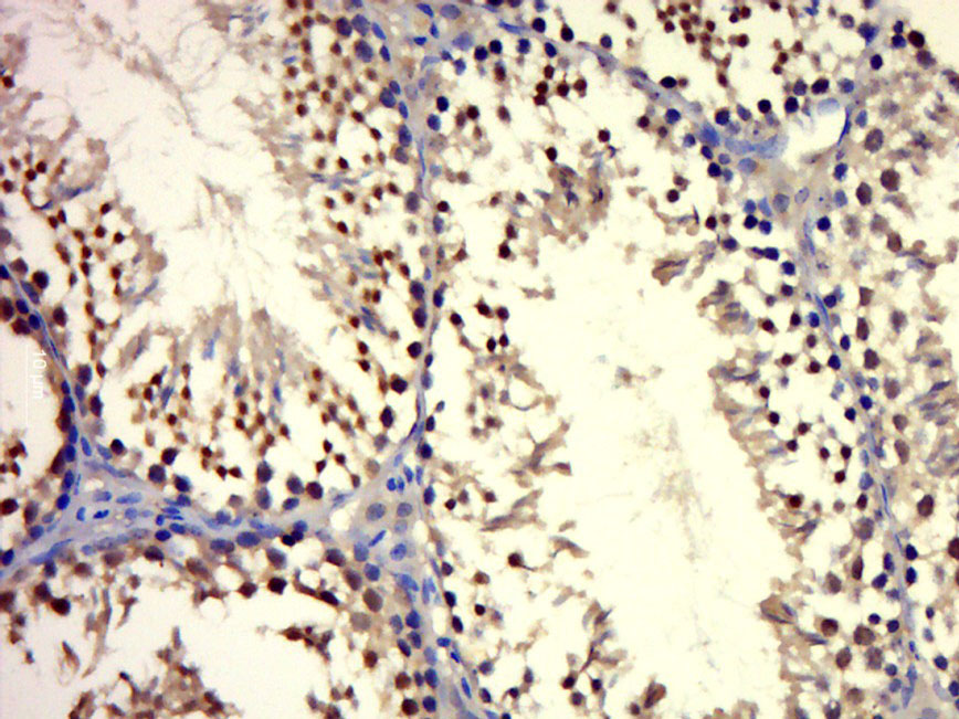
- Xiao Y, Nabi G, Hao Y, et al. Hormonal Regulation of Testicular Development in the Finless Porpoise Neophocaena asiaeorientalis sunameri: Preliminary Evidence from Testicular Histology and Immunohistochemistry. Zool Stud. 2018,57:e41. doi: 10.6620/ZS.2018.57-41Read this paper
- Guo J, Hu J, Cao R, et al. Androgen Receptor Is Inactivated and Degraded in Bladder Cancer Cells by Phenyl Glucosamine via miR-449a Restoration. Med Sci Monit. 2018,24:2294-2301.Read this paper
- Huang DY, Zheng CC, Pan Q, et al. Oral exposure of low-dose bisphenol A promotes proliferation of dorsolateral prostate and induces epithelial-mesenchymal transition in aged rats. Sci Rep. 2018,8(1):490. doi: 10.1038/s41598-017-18869-8Read this paper
- Zhang Y, Shen Y, Cao B, et al. Elevated expression levels of androgen receptors and matrix metalloproteinase-2 and -9 in 30 cases of hepatocellular carcinoma compared with adjacent tissues as predictors of cancer invasion and staging. Exp Ther Med. 2015,9(3):905-908.Read this paper
- Peng QH, Yao XL, Wu QL, et al. Effects of extract of Buddleja officinalis eye drops on androgen receptors of lacrimal gland cells of castrated rats with dry eye. Int J Ophthalmol. 2010,3(1):43-8. doi: 10.3980/j.issn.2222-3959.2010.01.10Read this paper
Related products
Product group Antibodies
ApplicationsImmunoPrecipitation, Western Blot, ImmunoCytoChemistry, ImmunoHistoChemistry
ReactivityRat
TargetAR
- SizePrice
Product group Antibodies
Anti-Androgen Receptor Antibody130-10000
ApplicationsWestern Blot, ELISA
ReactivityHuman
TargetAR
- SizePrice
Product group Antibodies
References
ApplicationsImmunoFluorescence, ImmunoCytoChemistry, ImmunoHistoChemistry, ImmunoHistoChemistry Frozen, ImmunoHistoChemistry Paraffin
ReactivityHuman, Mouse, Rat
TargetAR
- SizePrice

Product group Antibodies
Anti-AR AntibodyAMAB91547
ApplicationsWestern Blot, ImmunoHistoChemistry
ReactivityHuman
TargetAR
- SizePrice
Product group Antibodies

ApplicationsWestern Blot, ELISA
- SizePrice
Product group Antibodies
ApplicationsWestern Blot, ELISA
ReactivityHuman, Mouse, Rat
- SizePrice
Product group Antibodies

ApplicationsImmunoFluorescence, Western Blot, ChIP Chromatin ImmunoPrecipitation, ELISA, ImmunoHistoChemistry
ReactivityBovine, Canine, Human, Mouse, Porcine, Rat
TargetAR
- SizePrice
Product group Antibodies
References
ApplicationsImmunoFluorescence, Western Blot, ImmunoCytoChemistry, ImmunoHistoChemistry, ImmunoHistoChemistry Paraffin
ReactivityHuman, Mouse
TargetAR
- SizePrice