


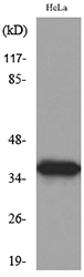
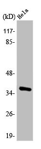
Figure 1. Western blot analysis of Annexin A2/ANXA2 using anti-Annexin A2/ANXA2 antibody (A00868-1). Electrophoresis was performed on a 5-20% SDS-PAGE gel at 70V (Stacking gel) / 90V (Resolving gel) for 2-3 hours. The sample well of each lane was loaded with 30 ug of sample under reducing conditions. Lane 1: human Hela whole cell lysates, Lane 2: human A549 whole cell lysates, Lane 3: human U251 whole cell lysates, Lane 4: human placenta tissue lysates, Lane 5: rat kidney tissue lysates, Lane 6: rat C6 whole cell lysates, Lane 7: mouse kidney tissue lysates, Lane 8: mouse NIH/3T3 whole cell lysates. After electrophoresis, proteins were transferred to a nitrocellulose membrane at 150 mA for 50-90 minutes. Blocked the membrane with 5% non-fat milk/TBS for 1.5 hour at RT. The membrane was incubated with rabbit anti-Annexin A2/ANXA2 antigen affinity purified polyclonal antibody (Catalog # A00868-1) at 0.5 microg/mL overnight at 4°C, then washed with TBS-0.1%Tween 3 times with 5 minutes each and probed with a goat anti-rabbit IgG-HRP secondary antibody at a dilution of 1:5000 for 1.5 hour at RT. The signal is developed using an Enhanced Chemiluminescent detection (ECL) kit (Catalog # EK1002) with Tanon 5200 system. A specific band was detected for Annexin A2/ANXA2 at approximately 36 kDa. The expected band size for Annexin A2/ANXA2 is at 39 kDa.
. Overlay histogram showing Hela cells stained with A00868-1 (Blue line). The cells were fixed with 4% paraformaldehyde and blocked with 10% normal goat serum. And then incubated with rabbit anti-Annexin A2/ANXA2 Antibody (A00868-1, 1 microg/1x106 cells) for 30 min at 20°C. DyLight®488 conjugated goat anti-rabbit IgG (BA1127, 5-10 microg/1x106 cells) was used as secondary antibody for 30 minutes at 20°C. Isotype control antibody (Green line) was rabbit IgG (1 microg/1x106) used under the same conditions. Unlabelled sample without incubation with primary antibody and secondary antibody (Red line) was used as a blank control.")
Figure 1. Western blot analysis of Annexin A2/ANXA2 using anti-Annexin A2/ANXA2 antibody (A00868-1). Electrophoresis was performed on a 5-20% SDS-PAGE gel at 70V (Stacking gel) / 90V (Resolving gel) for 2-3 hours. The sample well of each lane was loaded with 30 ug of sample under reducing conditions. Lane 1: human Hela whole cell lysates, Lane 2: human A549 whole cell lysates, Lane 3: human U251 whole cell lysates, Lane 4: human placenta tissue lysates, Lane 5: rat kidney tissue lysates, Lane 6: rat C6 whole cell lysates, Lane 7: mouse kidney tissue lysates, Lane 8: mouse NIH/3T3 whole cell lysates. After electrophoresis, proteins were transferred to a nitrocellulose membrane at 150 mA for 50-90 minutes. Blocked the membrane with 5% non-fat milk/TBS for 1.5 hour at RT. The membrane was incubated with rabbit anti-Annexin A2/ANXA2 antigen affinity purified polyclonal antibody (Catalog # A00868-1) at 0.5 microg/mL overnight at 4°C, then washed with TBS-0.1%Tween 3 times with 5 minutes each and probed with a goat anti-rabbit IgG-HRP secondary antibody at a dilution of 1:5000 for 1.5 hour at RT. The signal is developed using an Enhanced Chemiluminescent detection (ECL) kit (Catalog # EK1002) with Tanon 5200 system. A specific band was detected for Annexin A2/ANXA2 at approximately 36 kDa. The expected band size for Annexin A2/ANXA2 is at 39 kDa.
Anti-Annexin A2/ANXA2 Antibody Picoband(r)
A00868-1-PE
ApplicationsFlow Cytometry, Western Blot
Product group Antibodies
ReactivityHuman, Mouse, Rat
TargetANXA2
Overview
- SupplierBoster Bio
- Product NameAnti-Annexin A2/ANXA2 Antibody Picoband(r)
- Delivery Days Customer9
- ApplicationsFlow Cytometry, Western Blot
- CertificationResearch Use Only
- ClonalityPolyclonal
- Concentration500 ug/ml
- ConjugateRPE
- Gene ID302
- Target nameANXA2
- Target descriptionannexin A2
- Target synonymsANX2, ANX2L4, CAL1H, HEL-S-270, LIP2, LPC2, LPC2D, P36, PAP-IV, annexin A2, annexin II, annexin-2, calpactin I heavy chain, calpactin I heavy polypeptide, calpactin-1 heavy chain, chromobindin 8, epididymis secretory protein Li 270, epididymis secretory sperm binding protein, lipocortin II, placental anticoagulant protein IV, protein I
- HostRabbit
- Protein IDP07355
- Protein NameAnnexin A2
- Scientific DescriptionBoster Bio Anti-Annexin A2/ANXA2 Antibody Picoband® catalog # A00868-1. Tested in WB, Flow Cytometry applications. This antibody reacts with Human, Mouse, Rat. The brand Picoband indicates this is a premium antibody that guarantees superior quality, high affinity, and strong signals with minimal background in Western blot applications. Only our best-performing antibodies are designated as Picoband, ensuring unmatched performance.
- ReactivityHuman, Mouse, Rat
- Storage Instruction-20°C,2°C to 8°C
- UNSPSC12352203
Related products
Product group Antibodies
Anti-ANXA2 Antibody144-12397
ApplicationsImmunoFluorescence, Western Blot, ImmunoHistoChemistry
ReactivityHuman, Mouse, Rat
TargetANXA2
- SizePrice
Product group Antibodies

ApplicationsWestern Blot, ELISA
ReactivityBovine, Human, Mouse, Porcine, Rat
TargetANXA2
- SizePrice
Product group Antibodies
References
Annexin II antibody [N1], N-termGTX100046
ApplicationsImmunoFluorescence, ImmunoPrecipitation, Western Blot, ImmunoCytoChemistry, ImmunoHistoChemistry, ImmunoHistoChemistry Paraffin
ReactivityHuman
TargetANXA2
- SizePrice
Product group Antibodies
Anxa2 Polyclonal AntibodyCAC07082
ApplicationsImmunoFluorescence, ImmunoPrecipitation, Western Blot, ELISA, ImmunoHistoChemistry
ReactivityMouse
TargetANXA2
- SizePrice
Product group Antibodies
Anti-ANXA2 AntibodyA97716
ApplicationsWestern Blot, ELISA
ReactivityHuman, Mouse, Rat
- SizePrice
Product group Antibodies
Anti-Annexin A2/ANXA2 Antibody Picoband(r)A00868-1-CARRIER-FREE
ApplicationsFlow Cytometry, Western Blot
ReactivityHuman, Mouse, Rat
TargetANXA2
- SizePrice
Product group Antibodies
ANXA2 AntibodyCSB-PA000879
ApplicationsWestern Blot, ELISA, ImmunoHistoChemistry
ReactivityHuman, Mouse, Rat
TargetANXA2
- SizePrice

Product group Antibodies
Annexin A2 (2F4) Recombinant AntibodyBSM-54021R
ApplicationsFlow Cytometry, ImmunoFluorescence, Western Blot, ImmunoCytoChemistry, ImmunoHistoChemistry, ImmunoHistoChemistry Paraffin
ReactivityHuman
TargetANXA2
- SizePrice