


Figure 1. Western blot analysis of AP2 alpha using anti-AP2 alpha antibody (PB9118). Electrophoresis was performed on a 5-20% SDS-PAGE gel at 70V (Stacking gel) / 90V (Resolving gel) for 2-3 hours. Lane 1: recombinant human AP2A protein 0.5 ng. After electrophoresis, proteins were transferred to a nitrocellulose membrane at 150 mA for 50-90 minutes. Blocked the membrane with 5% non-fat milk/TBS for 1.5 hour at RT. The membrane was incubated with rabbit anti-AP2 alpha antigen affinity purified polyclonal antibody (Catalog # PB9118) at 0.5 microg/mL overnight at 4°C, then washed with TBS-0.1%Tween 3 times with 5 minutes each and probed with a goat anti-rabbit IgG-HRP secondary antibody at a dilution of 1:5000 for 1.5 hour at RT. The signal is developed using an Enhanced Chemiluminescent detection (ECL) kit (Catalog # EK1002) with Tanon 5200 system. A specific band was detected for AP2 alpha at approximately 38 kDa. The expected band size for AP2 alpha is at 38 kDa.
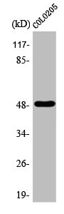
. Electrophoresis was performed on a 5-20% SDS-PAGE gel at 70V (Stacking gel) / 90V (Resolving gel) for 2-3 hours. The sample well of each lane was loaded with 30 ug of sample under reducing conditions. Lane 1: rat spleen tissue lysates. After electrophoresis, proteins were transferred to a nitrocellulose membrane at 150 mA for 50-90 minutes. Blocked the membrane with 5% non-fat milk/TBS for 1.5 hour at RT. The membrane was incubated with rabbit anti-AP2 alpha antigen affinity purified polyclonal antibody (Catalog # PB9118) at 0.5 microg/mL overnight at 4°C, then washed with TBS-0.1%Tween 3 times with 5 minutes each and probed with a goat anti-rabbit IgG-HRP secondary antibody at a dilution of 1:5000 for 1.5 hour at RT. The signal is developed using an Enhanced Chemiluminescent detection (ECL) kit (Catalog # EK1002) with Tanon 5200 system. A specific band was detected for AP2 alpha at approximately 48 kDa. The expected band size for AP2 alpha is at 48 kDa.")
Figure 1. Western blot analysis of AP2 alpha using anti-AP2 alpha antibody (PB9118). Electrophoresis was performed on a 5-20% SDS-PAGE gel at 70V (Stacking gel) / 90V (Resolving gel) for 2-3 hours. Lane 1: recombinant human AP2A protein 0.5 ng. After electrophoresis, proteins were transferred to a nitrocellulose membrane at 150 mA for 50-90 minutes. Blocked the membrane with 5% non-fat milk/TBS for 1.5 hour at RT. The membrane was incubated with rabbit anti-AP2 alpha antigen affinity purified polyclonal antibody (Catalog # PB9118) at 0.5 microg/mL overnight at 4°C, then washed with TBS-0.1%Tween 3 times with 5 minutes each and probed with a goat anti-rabbit IgG-HRP secondary antibody at a dilution of 1:5000 for 1.5 hour at RT. The signal is developed using an Enhanced Chemiluminescent detection (ECL) kit (Catalog # EK1002) with Tanon 5200 system. A specific band was detected for AP2 alpha at approximately 38 kDa. The expected band size for AP2 alpha is at 38 kDa.
Anti-AP2 alpha/TFAP2A Antibody Picoband(r)
PB9118-CARRIER-FREE
ApplicationsWestern Blot
Product group Antibodies
ReactivityHuman, Rat
TargetTFAP2A
Overview
- SupplierBoster Bio
- Product NameAnti-AP2 alpha/TFAP2A Antibody Picoband(r)
- Delivery Days Customer9
- Application Supplier NoteWB: The detection limit for AP2 alpha is approximately 0.25ng/lane under reducing conditions. Tested Species: In-house tested species with positive results. Other applications have not been tested. Optimal dilutions should be determined by end users.
- ApplicationsWestern Blot
- CertificationResearch Use Only
- ClonalityPolyclonal
- Concentration500 ug/ml
- Gene ID7020
- Target nameTFAP2A
- Target descriptiontranscription factor AP-2 alpha
- Target synonymsAP-2, AP-2alpha, AP2TF, BOFS, TFAP2, transcription factor AP-2-alpha, AP-2 transcription factor, activating enhancer-binding protein 2-alpha, activator protein 2, transcription factor AP-2 alpha (activating enhancer binding protein 2 alpha)
- HostRabbit
- IsotypeIgG
- Protein IDP05549
- Protein NameTranscription factor AP-2-alpha
- Scientific DescriptionBoster Bio Anti-AP2 alpha/TFAP2A Antibody Picoband® catalog # PB9118. Tested in WB applications. This antibody reacts with Human, Rat. The brand Picoband indicates this is a premium antibody that guarantees superior quality, high affinity, and strong signals with minimal background in Western blot applications. Only our best-performing antibodies are designated as Picoband, ensuring unmatched performance.
- ReactivityHuman, Rat
- Storage Instruction-20°C,2°C to 8°C
- UNSPSC12352203
Related products
Product group Antibodies
ReactivityHuman
TargetTFAP2A
- SizePrice
Product group Antibodies
TFAP2A/TFAP2B AntibodyCSB-PA000893
ApplicationsWestern Blot, ELISA, ImmunoHistoChemistry
ReactivityHuman, Mouse, Rat
TargetTFAP2A
- SizePrice

Product group Antibodies
Anti-TFAP2A AntibodyHPA028850
ApplicationsWestern Blot, ImmunoCytoChemistry, ImmunoHistoChemistry
ReactivityHuman, Rat
TargetTFAP2A
- SizePrice
Product group Antibodies

TFAP2A / AP-2 AntibodyLS-C496655
ApplicationsWestern Blot
ReactivityHuman, Mouse
TargetTFAP2A
- SizePrice
Product group Antibodies
AP2 alpha Polyclonal AntibodyBS-3569R
ApplicationsImmunoFluorescence, Western Blot, ELISA, ImmunoCytoChemistry, ImmunoHistoChemistry, ImmunoHistoChemistry Frozen, ImmunoHistoChemistry Paraffin
ReactivityBovine, Canine, Chicken, Equine, Human, Mouse, Porcine, Rabbit, Rat, Sheep
TargetTFAP2A
- SizePrice
![WB analysis of whole cell extracts (30 μg lysate) of PC-3 (Lane1), A-431 (Lane2), Mouse Placenta (Lane3) and RAW 264.7 (Lane5) using GTX15878 AP2 alpha antibody [3B5]. Dilution : 1:50-1:200](https://www.genetex.com/upload/website/prouct_img/normal/GTX15878/GTX15878_1549_WB_w_23060620_866.webp)
Product group Antibodies
AP2 alpha antibody [3B5]GTX15878
ApplicationsImmunoFluorescence, ImmunoPrecipitation, Western Blot, ImmunoCytoChemistry, ImmunoHistoChemistry, ImmunoHistoChemistry Paraffin
ReactivityChicken, Human, Mouse
TargetTFAP2A
- SizePrice
Product group Antibodies
Anti-AP2 alpha (N-term) Antibody102-23695
ApplicationsWestern Blot
TargetTFAP2A
- SizePrice