


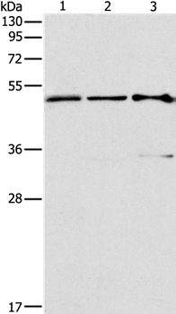
Figure 1. Western blot analysis of AP2 Gamma/TFAP2C using anti-AP2 Gamma/TFAP2C antibody (A01558-2). Electrophoresis was performed on a 5-20% SDS-PAGE gel at 70V (Stacking gel) / 90V (Resolving gel) for 2-3 hours. The sample well of each lane was loaded with 30 ug of sample under reducing conditions. Lane 1: human MCF-7 whole cell lysates. After electrophoresis, proteins were transferred to a nitrocellulose membrane at 150 mA for 50-90 minutes. Blocked the membrane with 5% non-fat milk/TBS for 1.5 hour at RT. The membrane was incubated with rabbit anti-AP2 Gamma/TFAP2C antigen affinity purified polyclonal antibody (Catalog # A01558-2) at 0.5 microg/mL overnight at 4°C, then washed with TBS-0.1%Tween 3 times with 5 minutes each and probed with a goat anti-rabbit IgG-HRP secondary antibody at a dilution of 1:5000 for 1.5 hour at RT. The signal is developed using an Enhanced Chemiluminescent detection (ECL) kit (Catalog # EK1002) with Tanon 5200 system. A specific band was detected for AP2 Gamma/TFAP2C at approximately 49 kDa. The expected band size for AP2 Gamma/TFAP2C is at 49 kDa.
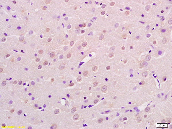
. AP2 Gamma/TFAP2C was detected in a paraffin-embedded section of human testis cancer tissue. Heat mediated antigen retrieval was performed in EDTA buffer (pH 8.0, epitope retrieval solution). The tissue section was blocked with 10% goat serum. The tissue section was then incubated with 2 microg/ml rabbit anti-AP2 Gamma/TFAP2C Antibody (A01558-2) overnight at 4°C. Biotinylated goat anti-rabbit IgG was used as secondary antibody and incubated for 30 minutes at 37°C. The tissue section was developed using Strepavidin-Biotin-Complex (SABC) (Catalog # SA1022) with DAB as the chromogen.")
. AP2 Gamma/TFAP2C was detected in a paraffin-embedded section of human testis cancer tissue. Heat mediated antigen retrieval was performed in EDTA buffer (pH 8.0, epitope retrieval solution). The tissue section was blocked with 10% goat serum. The tissue section was then incubated with 2 microg/ml rabbit anti-AP2 Gamma/TFAP2C Antibody (A01558-2) overnight at 4°C. Biotinylated goat anti-rabbit IgG was used as secondary antibody and incubated for 30 minutes at 37°C. The tissue section was developed using Strepavidin-Biotin-Complex (SABC) (Catalog # SA1022) with DAB as the chromogen.")
. AP2 Gamma/TFAP2C was detected in an immunocytochemical section of MCF-7 cells. Enzyme antigen retrieval was performed using IHC enzyme antigen retrieval reagent (AR0022) for 15 mins. The cells were blocked with 10% goat serum. And then incubated with 5 microg/mL rabbit anti-AP2 Gamma/TFAP2C Antibody (A01558-2) overnight at 4°C. DyLight®488 Conjugated Goat Anti-Rabbit IgG (BA1127) was used as secondary antibody at 1:100 dilution and incubated for 30 minutes at 37°C. The section was counterstained with DAPI. Visualize using a fluorescence microscope and filter sets appropriate for the label used.")
Figure 1. Western blot analysis of AP2 Gamma/TFAP2C using anti-AP2 Gamma/TFAP2C antibody (A01558-2). Electrophoresis was performed on a 5-20% SDS-PAGE gel at 70V (Stacking gel) / 90V (Resolving gel) for 2-3 hours. The sample well of each lane was loaded with 30 ug of sample under reducing conditions. Lane 1: human MCF-7 whole cell lysates. After electrophoresis, proteins were transferred to a nitrocellulose membrane at 150 mA for 50-90 minutes. Blocked the membrane with 5% non-fat milk/TBS for 1.5 hour at RT. The membrane was incubated with rabbit anti-AP2 Gamma/TFAP2C antigen affinity purified polyclonal antibody (Catalog # A01558-2) at 0.5 microg/mL overnight at 4°C, then washed with TBS-0.1%Tween 3 times with 5 minutes each and probed with a goat anti-rabbit IgG-HRP secondary antibody at a dilution of 1:5000 for 1.5 hour at RT. The signal is developed using an Enhanced Chemiluminescent detection (ECL) kit (Catalog # EK1002) with Tanon 5200 system. A specific band was detected for AP2 Gamma/TFAP2C at approximately 49 kDa. The expected band size for AP2 Gamma/TFAP2C is at 49 kDa.
Anti-AP2 gamma/TFAP2C Antibody Picoband(r)
A01558-2-CARRIER-FREE
ApplicationsImmunoFluorescence, Western Blot, ELISA, ImmunoCytoChemistry, ImmunoHistoChemistry
Product group Antibodies
ReactivityHuman
TargetTFAP2C
Overview
- SupplierBoster Bio
- Product NameAnti-AP2 gamma/TFAP2C Antibody Picoband(r)
- Delivery Days Customer9
- ApplicationsImmunoFluorescence, Western Blot, ELISA, ImmunoCytoChemistry, ImmunoHistoChemistry
- CertificationResearch Use Only
- ClonalityPolyclonal
- Concentration500 ug/ml
- Gene ID7022
- Target nameTFAP2C
- Target descriptiontranscription factor AP-2 gamma
- Target synonymsAP2-GAMMA, ERF1, TFAP2G, hAP-2g, transcription factor AP-2 gamma, activating enhancer-binding protein 2 gamma, estrogen receptor factor 1, transcription factor ERF-1
- HostRabbit
- IsotypeIgG
- Protein IDQ92754
- Protein NameTranscription factor AP-2 gamma
- Scientific DescriptionBoster Bio Anti-AP2 gamma/TFAP2C Antibody Picoband® catalog # A01558-2. Tested in ELISA, IF, IHC, ICC, WB applications. This antibody reacts with Human. The brand Picoband indicates this is a premium antibody that guarantees superior quality, high affinity, and strong signals with minimal background in Western blot applications. Only our best-performing antibodies are designated as Picoband, ensuring unmatched performance.
- ReactivityHuman
- Storage Instruction-20°C,2°C to 8°C
- UNSPSC12352203
Related products

Product group Antibodies
Anti-TFAP2C Antibody144-07739
ApplicationsWestern Blot
ReactivityHuman, Mouse, Rat
TargetTFAP2C
- SizePrice
Product group Antibodies
AP2 gamma Polyclonal AntibodyBS-6694R
ApplicationsImmunoFluorescence, Western Blot, ELISA, ImmunoCytoChemistry, ImmunoHistoChemistry, ImmunoHistoChemistry Frozen, ImmunoHistoChemistry Paraffin
ReactivityBovine, Canine, Human, Mouse, Rabbit, Rat
TargetTFAP2C
- SizePrice
Product group Antibodies
TFAP2C AntibodyCSB-PA000894
ApplicationsWestern Blot, ELISA, ImmunoHistoChemistry
ReactivityHuman, Mouse, Rat
TargetTFAP2C
- SizePrice
Product group Antibodies

Goat anti-AP-2 gammaEB06655
ApplicationsELISA, ImmunoHistoChemistry
ReactivityBovine, Canine, Human, Mouse, Porcine, Rat
TargetTFAP2C
- SizePrice
Product group Antibodies

TFAP2C / AP2 Gamma Antibody (AP)LS-C409290
ApplicationsWestern Blot, ImmunoHistoChemistry
ReactivityHuman, Mouse, Rat
TargetTFAP2C
- SizePrice

Product group Antibodies
Anti-TFAP2C AntibodyHPA055179
ApplicationsImmunoCytoChemistry, ImmunoHistoChemistry
ReactivityHuman
TargetTFAP2C
- SizePrice
![Non-transfected (–) and transfected (+) 293T whole cell extracts (30 μg) were separated by 10% SDS-PAGE, and the membranes were blotted with AP2 gamma antibody [N2C3] (GTX114833) diluted at 1:5000 and competitor's antibody (sc-8977) diluted at 1:1000. The HRP-conjugated anti-rabbit IgG antibody (GTX213110-01) was used to detect the primary antibody.](https://www.genetex.com/upload/website/prouct_img/normal/GTX114833/GTX114833_40548_20171117_WB_B_competitor_watermark_w_23060518_345.webp)
Product group Antibodies
AP2 gamma antibodyGTX114833
ApplicationsWestern Blot
ReactivityHuman
TargetTFAP2C
- SizePrice
Product group Antibodies
TargetTFAP2C
- SizePrice