


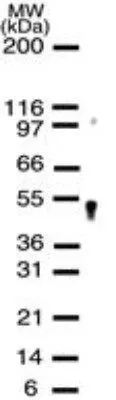
Figure 1. Western blot analysis of APEX2 using anti-APEX2 antibody (A07203-1). Electrophoresis was performed on a 5-20% SDS-PAGE gel at 70V (Stacking gel) / 90V (Resolving gel) for 2-3 hours. The sample well of each lane was loaded with 30 ug of sample under reducing conditions. Lane 1: human SH-SY5Y whole cell lysates, Lane 2: human 293T whole cell lysates. After electrophoresis, proteins were transferred to a nitrocellulose membrane at 150 mA for 50-90 minutes. Blocked the membrane with 5% non-fat milk/TBS for 1.5 hour at RT. The membrane was incubated with rabbit anti-APEX2 antigen affinity purified polyclonal antibody (Catalog # A07203-1) at 0.5 microg/mL overnight at 4°C, then washed with TBS-0.1%Tween 3 times with 5 minutes each and probed with a goat anti-rabbit IgG-HRP secondary antibody at a dilution of 1:5000 for 1.5 hour at RT. The signal is developed using an Enhanced Chemiluminescent detection (ECL) kit (Catalog # EK1002) with Tanon 5200 system. A specific band was detected for APEX2 at approximately 57 kDa. The expected band size for APEX2 is at 57 kDa.
. Overlay histogram showing HL-60 cells stained with A07203-1 (Blue line). To facilitate intracellular staining, cells were fixed with 4% paraformaldehyde and permeabilized with permeabilization buffer. The cells were blocked with 10% normal goat serum. And then incubated with rabbit anti-APEX2 Antibody (A07203-1, 1 microg/1x106 cells) for 30 min at 20°C. DyLight®488 conjugated goat anti-rabbit IgG (BA1127, 5-10 microg/1x106 cells) was used as secondary antibody for 30 minutes at 20°C. Isotype control antibody (Green line) was rabbit IgG (1 microg/1x106) used under the same conditions. Unlabelled sample (Red line) was also used as a control.")
Figure 1. Western blot analysis of APEX2 using anti-APEX2 antibody (A07203-1). Electrophoresis was performed on a 5-20% SDS-PAGE gel at 70V (Stacking gel) / 90V (Resolving gel) for 2-3 hours. The sample well of each lane was loaded with 30 ug of sample under reducing conditions. Lane 1: human SH-SY5Y whole cell lysates, Lane 2: human 293T whole cell lysates. After electrophoresis, proteins were transferred to a nitrocellulose membrane at 150 mA for 50-90 minutes. Blocked the membrane with 5% non-fat milk/TBS for 1.5 hour at RT. The membrane was incubated with rabbit anti-APEX2 antigen affinity purified polyclonal antibody (Catalog # A07203-1) at 0.5 microg/mL overnight at 4°C, then washed with TBS-0.1%Tween 3 times with 5 minutes each and probed with a goat anti-rabbit IgG-HRP secondary antibody at a dilution of 1:5000 for 1.5 hour at RT. The signal is developed using an Enhanced Chemiluminescent detection (ECL) kit (Catalog # EK1002) with Tanon 5200 system. A specific band was detected for APEX2 at approximately 57 kDa. The expected band size for APEX2 is at 57 kDa.
Anti-APEX2 Antibody Picoband(r)
A07203-1-CARRIER-FREE
ApplicationsFlow Cytometry, Western Blot, ELISA
Product group Antibodies
ReactivityHuman
TargetAPEX2
Overview
- SupplierBoster Bio
- Product NameAnti-APEX2 Antibody Picoband(r)
- Delivery Days Customer9
- Application Supplier NoteTested Species: In-house tested species with positive results. Predicted Species: Species predicted to be fit for the product based on sequence similarities. Other applications have not been tested. Optimal dilutions should be determined by end users.
- ApplicationsFlow Cytometry, Western Blot, ELISA
- CertificationResearch Use Only
- ClonalityPolyclonal
- Concentration500 ug/ml
- Gene ID27301
- Target nameAPEX2
- Target descriptionapurinic/apyrimidinic endodeoxyribonuclease 2
- Target synonymsAPE2, APEXL2, XTH2, ZGRF2, DNA-(apurinic or apyrimidinic site) endonuclease 2, AP endonuclease 2, AP endonuclease XTH2, APEX nuclease (apurinic/apyrimidinic endonuclease) 2, DNA-(apurinic or apyrimidinic site) lyase 2, apurinic/apyrimidinic endonuclease-like 2, zinc finger, GRF-type containing 2
- HostRabbit
- IsotypeIgG
- Protein IDQ9UBZ4
- Protein NameDNA-(apurinic or apyrimidinic site) endonuclease 2
- Scientific DescriptionBoster Bio Anti-APEX2 Antibody Picoband® catalog # A07203-1. Tested in ELISA, Flow Cytometry, WB applications. This antibody reacts with Human. The brand Picoband indicates this is a premium antibody that guarantees superior quality, high affinity, and strong signals with minimal background in Western blot applications. Only our best-performing antibodies are designated as Picoband, ensuring unmatched performance.
- ReactivityHuman
- Storage Instruction-20°C,2°C to 8°C
- UNSPSC12352203
Related products
Product group Antibodies

APEX2 AntibodyLS-C662254
ApplicationsWestern Blot
ReactivityHuman
TargetAPEX2
- SizePrice

Product group Antibodies
APEX2 Polyclonal AntibodyBS-6587R
ApplicationsWestern Blot, ELISA
ReactivityHuman, Mouse, Rat
TargetAPEX2
- SizePrice
Product group Antibodies
APEX2 antibodyGTX13691
ApplicationsWestern Blot
ReactivityHuman
TargetAPEX2
- SizePrice

Product group Antibodies
Anti-APEX2 AntibodyHPA048577
ApplicationsImmunoCytoChemistry
ReactivityHuman
TargetAPEX2
- SizePrice
Product group Antibodies
Anti-APEX2Y058378
ApplicationsELISA, ImmunoHistoChemistry
ReactivityHuman
- SizePrice