


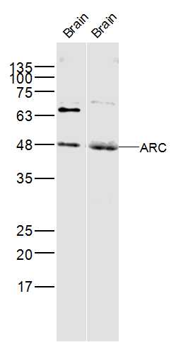
Figure 1. Western blot analysis of Arc using anti-Arc antibody (PB9753). Electrophoresis was performed on a 5-20% SDS-PAGE gel at 70V (Stacking gel) / 90V (Resolving gel) for 2-3 hours. The sample well of each lane was loaded with 30 ug of sample under reducing conditions. Lane 1: human U20S whole cell lysates, Lane 2: human 293T whole cell lysates, Lane 3: human SH-SY5Y whole cell lysates, Lane 4: human SiHa whole cell lysates, Lane 5: rat brain tissue lysates, Lane 6: rat C6 whole cell lysates, Lane 7: mouse brain tissue lysates, Lane 8: mouse Neuro-2a whole cell lysates. After electrophoresis, proteins were transferred to a nitrocellulose membrane at 150 mA for 50-90 minutes. Blocked the membrane with 5% non-fat milk/TBS for 1.5 hour at RT. The membrane was incubated with rabbit anti-Arc antigen affinity purified polyclonal antibody (Catalog # PB9753) at 0.5 microg/mL overnight at 4°C, then washed with TBS-0.1%Tween 3 times with 5 minutes each and probed with a goat anti-rabbit IgG-HRP secondary antibody at a dilution of 1:5000 for 1.5 hour at RT. The signal is developed using an Enhanced Chemiluminescent detection (ECL) kit (Catalog # EK1002) with Tanon 5200 system. A specific band was detected for Arc at approximately 45 kDa. The expected band size for Arc is at 45 kDa.
Figure 1. Western blot analysis of Arc using anti-Arc antibody (PB9753). Electrophoresis was performed on a 5-20% SDS-PAGE gel at 70V (Stacking gel) / 90V (Resolving gel) for 2-3 hours. The sample well of each lane was loaded with 30 ug of sample under reducing conditions. Lane 1: human U20S whole cell lysates, Lane 2: human 293T whole cell lysates, Lane 3: human SH-SY5Y whole cell lysates, Lane 4: human SiHa whole cell lysates, Lane 5: rat brain tissue lysates, Lane 6: rat C6 whole cell lysates, Lane 7: mouse brain tissue lysates, Lane 8: mouse Neuro-2a whole cell lysates. After electrophoresis, proteins were transferred to a nitrocellulose membrane at 150 mA for 50-90 minutes. Blocked the membrane with 5% non-fat milk/TBS for 1.5 hour at RT. The membrane was incubated with rabbit anti-Arc antigen affinity purified polyclonal antibody (Catalog # PB9753) at 0.5 microg/mL overnight at 4°C, then washed with TBS-0.1%Tween 3 times with 5 minutes each and probed with a goat anti-rabbit IgG-HRP secondary antibody at a dilution of 1:5000 for 1.5 hour at RT. The signal is developed using an Enhanced Chemiluminescent detection (ECL) kit (Catalog # EK1002) with Tanon 5200 system. A specific band was detected for Arc at approximately 45 kDa. The expected band size for Arc is at 45 kDa.
Anti-Arc Picoband Antibody
PB9753
ApplicationsWestern Blot
Product group Antibodies
ReactivityHuman, Mouse, Rat
TargetARC
Overview
- SupplierBoster Bio
- Product NameAnti-Arc Picoband Antibody
- Delivery Days Customer9
- Application Supplier NoteTested Species: In-house tested species with positive results. Other applications have not been tested. Optimal dilutions should be determined by end users.
- ApplicationsWestern Blot
- CertificationResearch Use Only
- ClonalityPolyclonal
- Concentration500 ug/ml
- Gene ID23237
- Target nameARC
- Target descriptionactivity regulated cytoskeleton associated protein
- Target synonymsArg3.1, hArc, activity-regulated cytoskeleton-associated protein, ARC/ARG3.1, activity-regulated gene 3.1 protein homolog
- HostRabbit
- IsotypeIgG
- Protein IDQ7LC44
- Protein NameActivity-regulated cytoskeleton-associated protein
- Scientific DescriptionBoster Bio Anti-Arc Antibody Picoband® catalog # PB9753. Tested in WB applications. This antibody reacts with Human, Mouse, Rat. The brand Picoband indicates this is a premium antibody that guarantees superior quality, high affinity, and strong signals with minimal background in Western blot applications. Only our best-performing antibodies are designated as Picoband, ensuring unmatched performance.
- ReactivityHuman, Mouse, Rat
- Storage Instruction-20°C,2°C to 8°C
- UNSPSC12352203
References
- Li Y, Gong ZH, Sheng L, et al. Anti-apoptotic effects of a calpain inhibitor on cardiomyocytes in a canine rapid atrial fibrillation model. Cardiovasc Drugs Ther. 2009,23(5):361-8. doi: 10.1007/s10557-009-6199-yRead this paper
Datasheet
MSDS
Related products
Product group Antibodies
ApplicationsImmunoPrecipitation, Western Blot, ImmunoCytoChemistry, ImmunoHistoChemistry
TargetARC
- SizePrice

Product group Antibodies
Anti-Arc AntibodyA10637
ApplicationsWestern Blot
ReactivityHuman, Mouse
- SizePrice
Product group Antibodies
ARC Polyclonal AntibodyBS-0385R
ApplicationsFlow Cytometry, Western Blot, ELISA
ReactivityBovine, Equine, Human, Mouse, Rat
TargetARC
- SizePrice
Product group Antibodies
Anti-ARC Antibody144-66726
ApplicationsWestern Blot
ReactivityHuman, Mouse
TargetARC
- SizePrice
Product group Antibodies
ARC antibodyGTX04096
ApplicationsImmunoFluorescence, Western Blot, ImmunoCytoChemistry, ImmunoHistoChemistry, ImmunoHistoChemistry Paraffin
ReactivityHuman, Mouse, Rat
TargetARC
- SizePrice
Product group Antibodies

ARC / Arg3.1 AntibodyLS-C401272
ApplicationsELISA, ImmunoHistoChemistry
ReactivityHuman
TargetARC
- SizePrice

Product group Antibodies
Anti-ARC AntibodyHPA056430
ApplicationsImmunoCytoChemistry
ReactivityHuman
TargetARC
- SizePrice
Product group Antibodies
ARC AntibodyCSB-PA698018
ApplicationsWestern Blot, ELISA, ImmunoHistoChemistry
ReactivityHuman, Mouse, Rat
TargetARC
- SizePrice
Product group Antibodies
Anti-ARCY058421
ApplicationsWestern Blot, ImmunoHistoChemistry
ReactivityHuman, Mouse
- SizePrice