

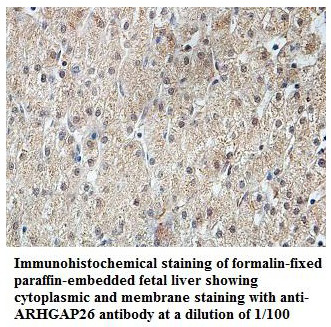

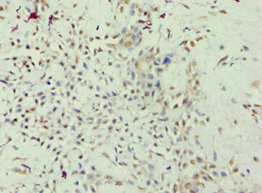
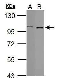
Anti-ARHGAP26 Antibody
A47443
ApplicationsWestern Blot, ELISA, ImmunoHistoChemistry
Product group Antibodies
ReactivityHuman, Mouse, Rat
Overview
- SupplierAntibodies.com
- Product NameAnti-ARHGAP26 Antibody
- Delivery Days Customer7
- ApplicationsWestern Blot, ELISA, ImmunoHistoChemistry
- CertificationResearch Use Only
- ClonalityPolyclonal
- Concentration1.0 mg/ml
- ConjugateUnconjugated
- HostRabbit
- Scientific DescriptionRabbit polyclonal antibody to ARHGAP26
- ReactivityHuman, Mouse, Rat
- UNSPSC12352203
Related products

Product group Antibodies
Anti-GRAF/ARHGAP26 Antibody Picoband(r)A08027-1-CARRIER-FREE
ApplicationsWestern Blot, ELISA
ReactivityHuman, Mouse, Rat
TargetARHGAP26
- SizePrice
Product group Antibodies

Goat anti-GRAF / ARHGAP26EB06142
ApplicationsWestern Blot, ELISA, ImmunoHistoChemistry
ReactivityBovine, Canine, Human, Mouse, Rat
TargetARHGAP26
- SizePrice

Product group Antibodies
Anti-ARHGAP26 AntibodyHPA035106
ApplicationsWestern Blot, ImmunoCytoChemistry, ImmunoHistoChemistry
ReactivityHuman
TargetARHGAP26
- SizePrice
Product group Antibodies

ARHGAP26 / GRAF Antibody (aa625-814)LS-C380287
ApplicationsELISA, ImmunoHistoChemistry, ImmunoHistoChemistry Paraffin
ReactivityHuman
TargetARHGAP26
- SizePrice
Product group Antibodies
ARHGAP26 AntibodyCSB-PA892479ESR1HU
ApplicationsELISA, ImmunoHistoChemistry
ReactivityHuman
TargetARHGAP26
- SizePrice
Product group Antibodies
Arhgap26 Polyclonal AntibodyCAC10646
ApplicationsELISA, ImmunoHistoChemistry
TargetARHGAP26
- SizePrice
Product group Antibodies
GRAF antibody [C3], C-termGTX107991
ApplicationsWestern Blot
ReactivityHuman, Mouse
TargetARHGAP26
- SizePrice
Product group Antibodies
Anti-ARHGAP26Y158233
ApplicationsWestern Blot, ELISA, ImmunoHistoChemistry
ReactivityHuman
- SizePrice
Product group Antibodies
GRAF Recombinant AntibodyBSM-62542R
ApplicationsWestern Blot
ReactivityHuman, Mouse, Rat
TargetARHGAP26
- SizePrice