


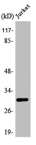
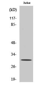
Figure 1. Western blot analysis of ATF5 using anti-ATF5 antibody (A02792-1). Electrophoresis was performed on a 5-20% SDS-PAGE gel at 70V (Stacking gel) / 90V (Resolving gel) for 2-3 hours. The sample well of each lane was loaded with 30 ug of sample under reducing conditions. Lane 1: human Jurkat whole cell lysates, Lane 2: human Hela whole cell lysates, Lane 3: human 293T whole cell lysates, Lane 4: human SH-SY5Y whole cell lysates, Lane 5: rat liver tissue lysates, Lane 6: rat RH35 whole cell lysates, Lane 7: mouse liver tissue lysates. After electrophoresis, proteins were transferred to a nitrocellulose membrane at 150 mA for 50-90 minutes. Blocked the membrane with 5% non-fat milk/TBS for 1.5 hour at RT. The membrane was incubated with rabbit anti-ATF5 antigen affinity purified polyclonal antibody (Catalog # A02792-1) at 0.5 microg/mL overnight at 4°C, then washed with TBS-0.1%Tween 3 times with 5 minutes each and probed with a goat anti-rabbit IgG-HRP secondary antibody at a dilution of 1:5000 for 1.5 hour at RT. The signal is developed using an Enhanced Chemiluminescent detection (ECL) kit (Catalog # EK1002) with Tanon 5200 system. A specific band was detected for ATF5 at approximately 37 kDa. The expected band size for ATF5 is at 31 kDa.
. Overlay histogram showing 293T cells stained with A02792-1 (Blue line). To facilitate intracellular staining, cells were fixed with 4% paraformaldehyde and permeabilized with permeabilization buffer. The cells were blocked with 10% normal goat serum. And then incubated with rabbit anti-ATF5 Antibody (A02792-1, 1 microg/1x106 cells) for 30 min at 20°C. DyLight®488 conjugated goat anti-rabbit IgG (BA1127, 5-10 microg/1x106 cells) was used as secondary antibody for 30 minutes at 20°C. Isotype control antibody (Green line) was rabbit IgG (1 microg/1x106) used under the same conditions. Unlabelled sample without incubation with primary antibody and secondary antibody (Red line) was used as a blank control.")
Figure 1. Western blot analysis of ATF5 using anti-ATF5 antibody (A02792-1). Electrophoresis was performed on a 5-20% SDS-PAGE gel at 70V (Stacking gel) / 90V (Resolving gel) for 2-3 hours. The sample well of each lane was loaded with 30 ug of sample under reducing conditions. Lane 1: human Jurkat whole cell lysates, Lane 2: human Hela whole cell lysates, Lane 3: human 293T whole cell lysates, Lane 4: human SH-SY5Y whole cell lysates, Lane 5: rat liver tissue lysates, Lane 6: rat RH35 whole cell lysates, Lane 7: mouse liver tissue lysates. After electrophoresis, proteins were transferred to a nitrocellulose membrane at 150 mA for 50-90 minutes. Blocked the membrane with 5% non-fat milk/TBS for 1.5 hour at RT. The membrane was incubated with rabbit anti-ATF5 antigen affinity purified polyclonal antibody (Catalog # A02792-1) at 0.5 microg/mL overnight at 4°C, then washed with TBS-0.1%Tween 3 times with 5 minutes each and probed with a goat anti-rabbit IgG-HRP secondary antibody at a dilution of 1:5000 for 1.5 hour at RT. The signal is developed using an Enhanced Chemiluminescent detection (ECL) kit (Catalog # EK1002) with Tanon 5200 system. A specific band was detected for ATF5 at approximately 37 kDa. The expected band size for ATF5 is at 31 kDa.
Anti-ATF5 Antibody Picoband(r)
A02792-1-CARRIER-FREE
ApplicationsFlow Cytometry, Western Blot, ELISA
Product group Antibodies
ReactivityHuman, Mouse, Rat
TargetATF5
Overview
- SupplierBoster Bio
- Product NameAnti-ATF5 Antibody Picoband(r)
- Delivery Days Customer9
- ApplicationsFlow Cytometry, Western Blot, ELISA
- CertificationResearch Use Only
- ClonalityPolyclonal
- Concentration500 ug/ml
- Gene ID22809
- Target nameATF5
- Target descriptionactivating transcription factor 5
- Target synonymsATFX, HMFN0395, cyclic AMP-dependent transcription factor ATF-5, cAMP-dependent transcription factor ATF-5, transcription factor ATFx
- HostRabbit
- IsotypeIgG
- Protein IDQ9Y2D1
- Protein NameCyclic AMP-dependent transcription factor ATF-5
- Scientific DescriptionBoster Bio Anti-ATF5 Antibody Picoband® catalog # A02792-1. Tested in WB, Flow Cytometry, ELISA applications. This antibody reacts with Human, Mouse, Rat. The brand Picoband indicates this is a premium antibody that guarantees superior quality, high affinity, and strong signals with minimal background in Western blot applications. Only our best-performing antibodies are designated as Picoband, ensuring unmatched performance.
- ReactivityHuman, Mouse, Rat
- Storage Instruction-20°C,2°C to 8°C
- UNSPSC12352203
Related products
Product group Antibodies
Anti-ATF5 AntibodyA98280
ApplicationsWestern Blot, ELISA
ReactivityHuman, Mouse, Rat
- SizePrice
Product group Antibodies
Anti-ATF5 Antibody144-65302
ApplicationsImmunoFluorescence, Western Blot
ReactivityHuman, Mouse, Rat
TargetATF5
- SizePrice
Product group Antibodies
ATF5 Recombinant Antibody, Biotin ConjugatedBSM-61324R-BIOTIN
ApplicationsImmunoPrecipitation, Western Blot, ImmunoHistoChemistry, ImmunoHistoChemistry Frozen, ImmunoHistoChemistry Paraffin
TargetATF5
- SizePrice
Product group Antibodies
ATF5 AntibodyCSB-PA000948
ApplicationsWestern Blot, ELISA, ImmunoHistoChemistry
ReactivityHuman, Mouse, Rat
TargetATF5
- SizePrice
Product group Antibodies

Goat anti-ATF5EB05246
ApplicationsWestern Blot, ELISA
ReactivityHuman, Mouse, Rat
TargetATF5
- SizePrice
Product group Antibodies
Atf5 Recombinant AntibodyCAC12001
ApplicationsWestern Blot, ELISA, ImmunoHistoChemistry
ReactivityRat
TargetATF5
- SizePrice
Product group Antibodies

ATF5 Antibody (acetylation site of K29.)LS-C380578
ApplicationsWestern Blot, ELISA
ReactivityHuman, Mouse, Rat
TargetATF5
- SizePrice
Product group Antibodies
Anti-ATF5 AntibodyHPA030187
ApplicationsWestern Blot, ChIP Chromatin ImmunoPrecipitation, ImmunoCytoChemistry, ImmunoHistoChemistry
ReactivityHuman
TargetATF5
- SizePrice
Product group Antibodies
ATF5 antibodyGTX34045
ApplicationsWestern Blot
ReactivityHuman
TargetATF5
- SizePrice