


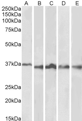
Figure 1. Western blot analysis of Aurora B using anti-Aurora B antibody (M00762-1). Electrophoresis was performed on a 5-20% SDS-PAGE gel at 70V (Stacking gel) / 90V (Resolving gel) for 2-3 hours. The sample well of each lane was loaded with 30 ug of sample under reducing conditions. Lane 1: human Hela whole cell lysates, Lane 2: human 293T whole cell lysates, Lane 3: human Jurkat whole cell lysates, Lane 4: human U251 whole cell lysates. After electrophoresis, proteins were transferred to a nitrocellulose membrane at 150 mA for 50-90 minutes. Blocked the membrane with 5% non-fat milk/TBS for 1.5 hour at RT. The membrane was incubated with rabbit anti-Aurora B antigen affinity purified monoclonal antibody (Catalog # M00762-1) at 1:1000 overnight at 4°C, then washed with TBS-0.1%Tween 3 times with 5 minutes each and probed with a goat anti-rabbit IgG-HRP secondary antibody at a dilution of 1:500 for 1.5 hour at RT. The signal is developed using an Enhanced Chemiluminescent detection (ECL) kit (Catalog # EK1002) with Tanon 5200 system. A specific band was detected for Aurora B at approximately 39 kDa. The expected band size for Aurora B is at 39 kDa.
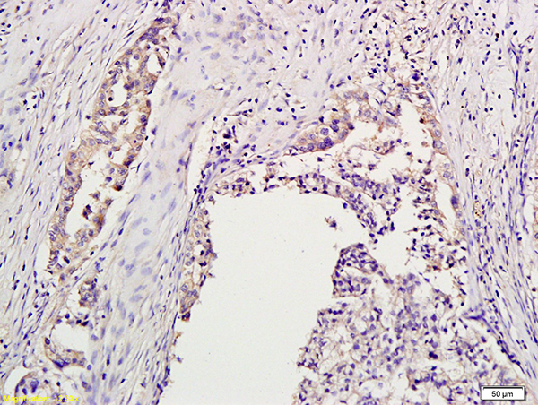
. AURKB was detected in a paraffin-embedded section of human bladder urothelial carcinoma tissue. Heat mediated antigen retrieval was performed in EDTA buffer (pH 8.0, epitope retrieval solution). The tissue section was blocked with 10% goat serum. The tissue section was then incubated with 1:50 rabbit anti-AURKB Antibody (M00762-1) overnight at 4°C. Peroxidase Conjugated Goat Anti-rabbit IgG was used as secondary antibody and incubated for 30 minutes at 37°C. The tissue section was developed using HRP Conjugated Rabbit IgG Super Vision Assay Kit (Catalog # SV0002) with DAB as the chromogen.")
. AURKB was detected in a paraffin-embedded section of human tonsil tissue. Heat mediated antigen retrieval was performed in EDTA buffer (pH 8.0, epitope retrieval solution). The tissue section was blocked with 10% goat serum. The tissue section was then incubated with 1:50 rabbit anti-AURKB Antibody (M00762-1) overnight at 4°C. Peroxidase Conjugated Goat Anti-rabbit IgG was used as secondary antibody and incubated for 30 minutes at 37°C. The tissue section was developed using HRP Conjugated Rabbit IgG Super Vision Assay Kit (Catalog # SV0002) with DAB as the chromogen.")
Figure 1. Western blot analysis of Aurora B using anti-Aurora B antibody (M00762-1). Electrophoresis was performed on a 5-20% SDS-PAGE gel at 70V (Stacking gel) / 90V (Resolving gel) for 2-3 hours. The sample well of each lane was loaded with 30 ug of sample under reducing conditions. Lane 1: human Hela whole cell lysates, Lane 2: human 293T whole cell lysates, Lane 3: human Jurkat whole cell lysates, Lane 4: human U251 whole cell lysates. After electrophoresis, proteins were transferred to a nitrocellulose membrane at 150 mA for 50-90 minutes. Blocked the membrane with 5% non-fat milk/TBS for 1.5 hour at RT. The membrane was incubated with rabbit anti-Aurora B antigen affinity purified monoclonal antibody (Catalog # M00762-1) at 1:1000 overnight at 4°C, then washed with TBS-0.1%Tween 3 times with 5 minutes each and probed with a goat anti-rabbit IgG-HRP secondary antibody at a dilution of 1:500 for 1.5 hour at RT. The signal is developed using an Enhanced Chemiluminescent detection (ECL) kit (Catalog # EK1002) with Tanon 5200 system. A specific band was detected for Aurora B at approximately 39 kDa. The expected band size for Aurora B is at 39 kDa.
Anti-Aurora B AURKB Rabbit Monoclonal Antibody
M00762-1
ApplicationsImmunoFluorescence, ImmunoPrecipitation, Western Blot, ImmunoCytoChemistry, ImmunoHistoChemistry
Product group Antibodies
ReactivityHuman
TargetAURKB
Overview
- SupplierBoster Bio
- Product NameAnti-Aurora B AURKB Rabbit Monoclonal Antibody
- Delivery Days Customer9
- ApplicationsImmunoFluorescence, ImmunoPrecipitation, Western Blot, ImmunoCytoChemistry, ImmunoHistoChemistry
- CertificationResearch Use Only
- ClonalityMonoclonal
- Clone IDFA-1
- Gene ID9212
- Target nameAURKB
- Target descriptionaurora kinase B
- Target synonymsAIK2, AIM-1, AIM1, ARK-2, ARK2, AurB, IPL1, PPP1R48, STK-1, STK12, STK5, aurkb-sv1, aurkb-sv2, aurora kinase B, aurora kinase B-Sv1, aurora kinase B-Sv2, aurora- and Ipl1-like midbody-associated protein 1, aurora- and Ipl1-like midbody-associated protein 1 homolog, aurora-1, aurora-B, aurora-related kinase 2, aurora/IPL1-related kinase 2, protein phosphatase 1, regulatory subunit 48, serine/threonine kinase 12, serine/threonine-protein kinase 12, serine/threonine-protein kinase 5, serine/threonine-protein kinase aurora-B
- HostRabbit
- IsotypeIgG
- Protein IDQ96GD4
- Protein NameAurora kinase B
- Scientific DescriptionBoster Bio Anti-Aurora B AURKB Rabbit Monoclonal Antibody catalog # M00762-1. Tested in WB, IHC, ICC/IF, IP applications. This antibody reacts with Human.
- ReactivityHuman
- Storage Instruction-20°C
- UNSPSC12352203
Datasheet
MSDS
Related products
Product group Antibodies
Anti-Aurora B AntibodyA84215
ApplicationsWestern Blot, ELISA
ReactivityHuman
- SizePrice
Product group Antibodies
References
Aurora B Polyclonal AntibodyBS-2445R
ApplicationsFlow Cytometry, Western Blot, ELISA, ImmunoHistoChemistry, ImmunoHistoChemistry Paraffin
ReactivityBovine, Equine, Human, Mouse, Porcine, Rabbit, Rat
TargetAURKB
- SizePrice
Product group Antibodies
AURKB AntibodyCSB-PA000926
ApplicationsImmunoFluorescence, Western Blot, ELISA, ImmunoHistoChemistry
ReactivityHuman, Monkey, Mouse, Rat
TargetAURKB
- SizePrice
Product group Antibodies

Goat anti-Aurora Kinase BEB07260
ApplicationsWestern Blot, ELISA
ReactivityCanine, Human, Mouse, Rat
TargetAURKB
- SizePrice
Product group Antibodies
Aurkb Polyclonal AntibodyCAC07483
ApplicationsImmunoFluorescence, ELISA, ImmunoHistoChemistry
TargetAURKB
- SizePrice
Product group Antibodies

AURKB / Aurora-B Antibody (N-Terminus)LS-C358921
ApplicationsImmunoFluorescence, ImmunoPrecipitation, Western Blot, ImmunoCytoChemistry, ImmunoHistoChemistry, ImmunoHistoChemistry Paraffin
ReactivityBovine, Human, Mouse, Porcine, Rat
TargetAURKB
- SizePrice
Product group Antibodies
Aurora B antibodyGTX130211
ApplicationsWestern Blot
ReactivityHuman
TargetAURKB
- SizePrice

Product group Antibodies
Anti-AURKB AntibodyHPA037708
ApplicationsImmunoCytoChemistry
ReactivityHuman
TargetAURKB
- SizePrice