


Figure 1. Western blot analysis of Bax using anti-Bax antibody (PA1013-1). Electrophoresis was performed on a 5-20% SDS-PAGE gel at 70V (Stacking gel) / 90V (Resolving gel) for 2-3 hours. The sample well of each lane was loaded with 50ug of sample under reducing conditions. Lane 1: Rat Testis Tissue Lysate Lane 2: Rat Kidney Tissue Lysate Lane 3: Rat Brain Tissue Lysate Lane 4: Rat Ovary Tissue Lysate Lane 5: HELA Cell Lysate Lane 6: MM231 Cell Lysate Lane 7: A549 Cell Lysate Lane 8: JURKAT Cell Lysate Lane 9: Human Placenta Tissue Lysate After Electrophoresis, proteins were transferred to a Nitrocellulose membrane at 150mA for 50-90 minutes. Blocked the membrane with 5% Non-fat Milk/ TBS for 1.5 hour at RT. The membrane was incubated with rabbit anti-Bax antigen affinity purified polyclonal antibody (Catalog # PA1013-1) at 0.5 microg/mL overnight at 4°C, then washed with TBS-0.1%Tween 3 times with 5 minutes each and probed with a goat anti-rabbit IgG-HRP secondary antibody at a dilution of 1:10000 for 1.5 hour at RT. The signal is developed using an Enhanced Chemiluminescent detection (ECL) kit (Catalog # EK1002) with Tanon 5200 system. A specific band was detected for Bax at approximately 21&28KD. The expected band size for Bax is at 21KD.
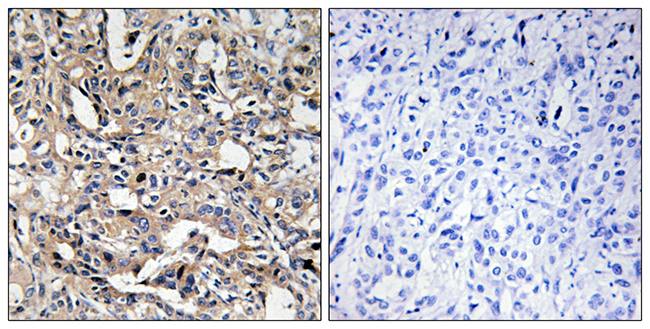
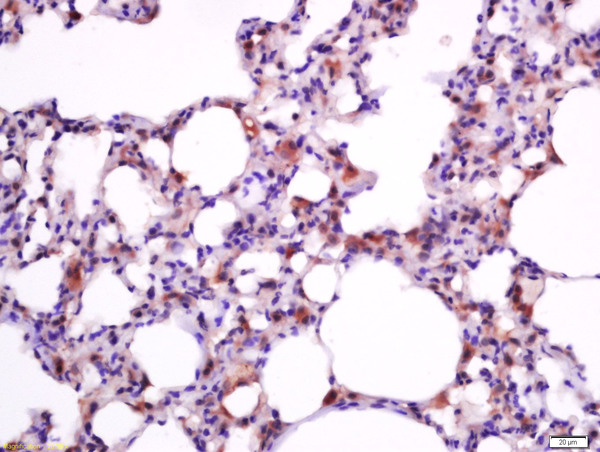
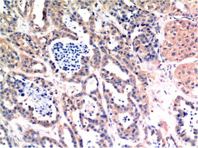
. Bax was detected in a paraffin-embedded section of human ovarian cancer tissue. Heat mediated antigen retrieval was performed in EDTA buffer (pH 8.0, epitope retrieval solution). The tissue section was blocked with 10% goat serum. The tissue section was then incubated with 2 microg/ml rabbit anti-Bax Antibody (PA1013-1) overnight at 4°C. Peroxidase Conjugated Goat Anti-rabbit IgG was used as secondary antibody and incubated for 30 minutes at 37°C. The tissue section was developed using HRP Conjugated Rabbit IgG Super Vision Assay Kit (Catalog # SV0002) with DAB as the chromogen.")
. Overlay histogram showing A431 cells stained with PA1013-1 (Blue line). To facilitate intracellular staining, cells were fixed with 4% paraformaldehyde and permeabilized with permeabilization buffer. The cells were blocked with 10% normal goat serum. And then incubated with rabbit anti-BAX Antibody (PA1013-1,1microg/1x106 cells) for 30 min at 20°C. DyLight®488 conjugated goat anti-rabbit IgG (BA1127, 5-10microg/1x106 cells) was used as secondary antibody for 30 minutes at 20°C. Isotype control antibody (Green line) was rabbit IgG (1microg/1x106) used under the same conditions. Unlabelled sample without incubation with primary antibody and secondary antibody (Red line) was used as a blank control.")
Figure 1. Western blot analysis of Bax using anti-Bax antibody (PA1013-1). Electrophoresis was performed on a 5-20% SDS-PAGE gel at 70V (Stacking gel) / 90V (Resolving gel) for 2-3 hours. The sample well of each lane was loaded with 50ug of sample under reducing conditions. Lane 1: Rat Testis Tissue Lysate Lane 2: Rat Kidney Tissue Lysate Lane 3: Rat Brain Tissue Lysate Lane 4: Rat Ovary Tissue Lysate Lane 5: HELA Cell Lysate Lane 6: MM231 Cell Lysate Lane 7: A549 Cell Lysate Lane 8: JURKAT Cell Lysate Lane 9: Human Placenta Tissue Lysate After Electrophoresis, proteins were transferred to a Nitrocellulose membrane at 150mA for 50-90 minutes. Blocked the membrane with 5% Non-fat Milk/ TBS for 1.5 hour at RT. The membrane was incubated with rabbit anti-Bax antigen affinity purified polyclonal antibody (Catalog # PA1013-1) at 0.5 microg/mL overnight at 4°C, then washed with TBS-0.1%Tween 3 times with 5 minutes each and probed with a goat anti-rabbit IgG-HRP secondary antibody at a dilution of 1:10000 for 1.5 hour at RT. The signal is developed using an Enhanced Chemiluminescent detection (ECL) kit (Catalog # EK1002) with Tanon 5200 system. A specific band was detected for Bax at approximately 21&28KD. The expected band size for Bax is at 21KD.
Anti-Apoptosis regulator BAX Bax Antibody Picoband(r)
PA1013-1
ApplicationsFlow Cytometry, Western Blot, ImmunoHistoChemistry
Product group Antibodies
ReactivityHamster, Human, Mouse, Rat
TargetBAX
Overview
- SupplierBoster Bio
- Product NameAnti-Apoptosis regulator BAX Bax Antibody Picoband(r)
- Delivery Days Customer9
- ApplicationsFlow Cytometry, Western Blot, ImmunoHistoChemistry
- Applications SupplierWB
- CertificationResearch Use Only
- ClonalityPolyclonal
- Concentration500 ug/ml
- Gene ID581
- Target nameBAX
- Target descriptionBCL2 associated X, apoptosis regulator
- Target synonymsBCL2L4, apoptosis regulator BAX, BCL2 associated X protein, BCL2-associated X protein omega, Baxdelta2(G8)-RFS protein, Baxdelta2G9, Baxdelta2G9omega, Baxdelta2omega, bcl-2-like protein 4, bcl2-L-4
- HostRabbit
- IsotypeIgG
- Protein IDQ07812
- Protein NameApoptosis regulator BAX
- Scientific DescriptionBoster Bio Anti-Apoptosis regulator BAX Bax Antibody catalog # PA1013-1. Tested in Flow Cytometry, IHC, WB applications. This antibody reacts with Human, Mouse, Rat. The brand Picoband indicates this is a premium antibody that guarantees superior quality, high affinity, and strong signals with minimal background in Western blot applications. Only our best-performing antibodies are designated as Picoband, ensuring unmatched performance.
- ReactivityHamster, Human, Mouse, Rat
- Reactivity SupplierHuman, Mouse, Rat, Hamster
- Storage Instruction-20°C,2°C to 8°C
- UNSPSC12352203
References
- Xu H, Zheng Y, Wu J, et al. circSORBS1 inhibits lung cancer progression by sponging miR-6779-5p and directly binding RUFY3 mRNA. J Transl Med. 2024,22(1):590. doi: 10.1186/s12967-024-05423-0Read this paper
- Duan P, Li B, Zhou Y, et al. ZBTB20 suppresses tumor growth in glioblastoma through activating the TET1/FAS/caspase‑3 pathway. Oncol Lett. 2024,28(2):358. doi: 10.3892/ol.2024.14491Read this paper
- Zhu Y, Zhang S, Shao Y, et al. Regulatory role of oxidative stress in retrorsine - Induced apoptosis and autophagy in primary rat hepatocytes. Ecotoxicol Environ Saf. 2024,279:116515. doi: 10.1016/j.ecoenv.2024.116515Read this paper
- Cai J, Lin Y, Zhou B, et al. SHARPIN contributes to sevoflurane-induced neonatal neurotoxicity through up-regulating HMGB1 to repress M2 like-macrophage polarization. Metab Brain Dis. 2024,39(5):841-853. doi: 10.1007/s11011-024-01355-2Read this paper
- Chen F, Yu X. Circ_0002331 Interacts with ELAVL1 to Improve ox-LDL-Induced Vascular Endothelial Cell Dysfunction via Regulating CCND2 mRNA Stability. Cardiovasc Toxicol. 2024,24(7):625-636. doi: 10.1007/s12012-024-09865-2Read this paper
- Zheng H, Liang G, Guan C, et al. Mitochondrial Fission in Nickel Nanoparticle-Induced Reproductive Toxicity: An In Vitro GC-1 Cell Study. Nanomaterials (Basel). 2024,14(8). doi: 10.3390/nano14080689Read this paper
- Wu J, Cui Y, Ding W, et al. The protective effect of Macrostemonoside T from Allium macrostemon Bunge against Isoproterenol-Induced myocardial injury via the PI3K/Akt/mTOR signaling pathway. Int Immunopharmacol. 2024,133:112086. doi: 10.1016/j.intimp.2024.112086Read this paper
- Li Y, Fan F, Liu Q. Cytisine-N-methylene-(5,7,4(')-trihydroxy)- isoflavone ameliorates ischemic stroke-induced brain injury in mouse by regulating the oxidative stress and BDNF-Trkb/Akt pathway. Eur J Pharmacol. 2024,974:176512. doi: 10.1016/j.ejphar.2024.176512Read this paper
- Xiao M, Pu X, Tong W, et al. Declined circular RNA mitofusin 2 constrains the deterioration of Wilms tumor via modulating microRNA-372-3p/transforming growth factor-β receptor type 2 axis. Cell Mol Biol (Noisy-le-grand). 2024,70(2):143-149. doi: 10.14715/cmb/2024.70.2.20Read this paper
- Wang H, Li C, Zhu L, et al. Adiponectin attenuates H2O2-induced apoptosis in chicken skeletal myoblasts through the lysosomal-mitochondrial axis. In Vitro Cell Dev Biol Anim. 2024,60(7):805-814. doi: 10.1007/s11626-024-00857-8Read this paper
Datasheet
MSDS
Related products
Product group Antibodies

Anti-Bax (active monomer) [6A7]Ab00120-1.1
ApplicationsImmunoPrecipitation, Western Blot, ImmunoHistoChemistry
ReactivityHuman, Mouse, Rat
TargetBAX
- SizePrice
Product group Antibodies
Anti-BAX AntibodyA101574
ApplicationsELISA, ImmunoHistoChemistry
ReactivityHuman
- SizePrice

Product group Antibodies
Anti-BAX AntibodyAMAB91490
ApplicationsWestern Blot, ImmunoHistoChemistry
ReactivityHuman
TargetBAX
- SizePrice

Product group Antibodies
Anti-Bax Antibody Picoband(r)A00183-CARRIER-FREE
ApplicationsFlow Cytometry, ImmunoFluorescence, Western Blot, ImmunoCytoChemistry, ImmunoHistoChemistry
ReactivityHuman, Mouse, Rat
TargetBAX
- SizePrice
Product group Antibodies
References
Bax Polyclonal AntibodyBS-0127R
ApplicationsFlow Cytometry, ImmunoFluorescence, ImmunoPrecipitation, Western Blot, ELISA, ImmunoCytoChemistry, ImmunoHistoChemistry, ImmunoHistoChemistry Frozen, ImmunoHistoChemistry Paraffin
ReactivityBovine, Canine, Human, Mouse, Porcine, Rabbit, Rat, Sheep
TargetBAX
- SizePrice
Product group Antibodies
BAX Monoclonal AntibodyCSB-MA183751
ApplicationsWestern Blot, ELISA, ImmunoHistoChemistry
ReactivityHuman, Mouse, Rat
TargetBAX
- SizePrice
Product group Antibodies
ApplicationsImmunoPrecipitation, Western Blot, ImmunoCytoChemistry, ImmunoHistoChemistry
ReactivityMouse, Porcine, Rat
TargetBAX
- SizePrice
Product group Antibodies

BAX Antibody (Preservative Free)LS-C149225
ApplicationsWestern Blot, ELISA
ReactivityHuman
TargetBAX
- SizePrice
![Various whole cell extracts (30 μg) were separated by 12% SDS-PAGE, and the membrane was blotted with Bax antibody [N1N2], N-term (GTX109683) diluted at 1:1000. The HRP-conjugated anti-rabbit IgG antibody (GTX213110-01) was used to detect the primary antibody.](https://www.genetex.com/upload/website/prouct_img/normal/GTX109683/GTX109683_44286_20210806_WB_R_w_23060500_215.webp)
Product group Antibodies
Bax antibody [N1N2], N-termGTX109683
ApplicationsImmunoFluorescence, ImmunoPrecipitation, Western Blot, ImmunoCytoChemistry, ImmunoHistoChemistry, ImmunoHistoChemistry Frozen, ImmunoHistoChemistry Paraffin
ReactivityHuman, Mouse, Rat
TargetBAX
- SizePrice