

Various whole cell extracts (30 μg) were separated by 12% SDS-PAGE, and the membrane was blotted with Bax antibody [N1N2], N-term (GTX109683) diluted at 1:1000. The HRP-conjugated anti-rabbit IgG antibody (GTX213110-01) was used to detect the primary antibody.
![Various whole cell extracts (30 μg) were separated by 12% SDS-PAGE, and the membrane was blotted with Bax antibody [N1N2], N-term (GTX109683) diluted at 1:1000. The HRP-conjugated anti-rabbit IgG antibody (GTX213110-01) was used to detect the primary antibody.](https://www.genetex.com/upload/website/prouct_img/normal/GTX109683/GTX109683_44286_20210806_WB_M_w_23060500_765.webp "Various whole cell extracts (30 μg) were separated by 12% SDS-PAGE, and the membrane was blotted with Bax antibody [N1N2], N-term (GTX109683) diluted at 1:1000. The HRP-conjugated anti-rabbit IgG antibody (GTX213110-01) was used to detect the primary antibody.")
![Bax antibody [N1N2], N-term detects Bax protein at cytoplasm, nucleus and mitochondria by immunofluorescent analysis. Sample: HeLa cells were fixed in 4% paraformaldehyde at RT for 15 min. Green: Bax protein stained by Bax antibody [N1N2], N-term (GTX109683) diluted at 1:400. Blue: Hoechst 33342 staining.](https://www.genetex.com/upload/website/prouct_img/normal/GTX109683/GTX109683_39988_20160525_IFA_w_23060500_584.webp "Bax antibody [N1N2], N-term detects Bax protein at cytoplasm, nucleus and mitochondria by immunofluorescent analysis. Sample: HeLa cells were fixed in 4% paraformaldehyde at RT for 15 min. Green: Bax protein stained by Bax antibody [N1N2], N-term (GTX109683) diluted at 1:400. Blue: Hoechst 33342 staining.")
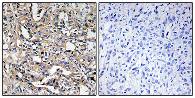
![Bax antibody [N1N2], N-term detects Bax protein at cytoplasm on human lung carcinoma by immunohistochemical analysis. Sample: Paraffin-embedded human lung carcinoma. Bax antibody [N1N2], N-term (GTX109683) diluted at 1:500.
Antigen Retrieval: Trilogy? (EDTA based, pH 8.0) buffer, 15min](https://www.genetex.com/upload/website/prouct_img/normal/GTX109683/GTX109683_39988_20141205_IHC_w_23060500_668.webp "Bax antibody [N1N2], N-term detects Bax protein at cytoplasm on human lung carcinoma by immunohistochemical analysis. Sample: Paraffin-embedded human lung carcinoma. Bax antibody [N1N2], N-term (GTX109683) diluted at 1:500.
Antigen Retrieval: Trilogy? (EDTA based, pH 8.0) buffer, 15min")
![Various whole cell extracts (30 μg) were separated by 12% SDS-PAGE, and the membrane was blotted with Bax antibody [N1N2], N-term (GTX109683) diluted at 1:1000. The HRP-conjugated anti-rabbit IgG antibody (GTX213110-01) was used to detect the primary antibody. Corresponding RNA expression data for the same cell lines are based on Human Protein Atlas program.](https://www.genetex.com/upload/website/prouct_img/normal/GTX109683/GTX109683_44286_20210416_WB_TPM_watermark_w_23060500_172.webp "Various whole cell extracts (30 μg) were separated by 12% SDS-PAGE, and the membrane was blotted with Bax antibody [N1N2], N-term (GTX109683) diluted at 1:1000. The HRP-conjugated anti-rabbit IgG antibody (GTX213110-01) was used to detect the primary antibody. Corresponding RNA expression data for the same cell lines are based on Human Protein Atlas program.")
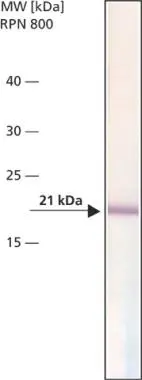
![Bax antibody [N1N2], N-term immunoprecipitates BCL2-associated X protein in IP experiments. IP samples: MCF-7 whole cell extract A. Control with 4 μg of preimmune Rabbit IgG B. Immunoprecipitation of BCL2-associated X protein by 4 μg Bax antibody [N1N2], N-term (GTX109683) 15 % SDS-PAGE The immunoprecipitated BCL2-associated X protein was detected by Bax antibody [N1N2], N-term (GTX109683) diluted at 1:500. [EasyBlot anti-rabbit IgG (GTX221666-01) was used as a secondary reagent]](https://www.genetex.com/upload/website/prouct_img/normal/GTX109683/GTX109683_39988_IP_w_23060500_274.webp "Bax antibody [N1N2], N-term immunoprecipitates BCL2-associated X protein in IP experiments. IP samples: MCF-7 whole cell extract A. Control with 4 μg of preimmune Rabbit IgG B. Immunoprecipitation of BCL2-associated X protein by 4 μg Bax antibody [N1N2], N-term (GTX109683) 15 % SDS-PAGE The immunoprecipitated BCL2-associated X protein was detected by Bax antibody [N1N2], N-term (GTX109683) diluted at 1:500. [EasyBlot anti-rabbit IgG (GTX221666-01) was used as a secondary reagent]")
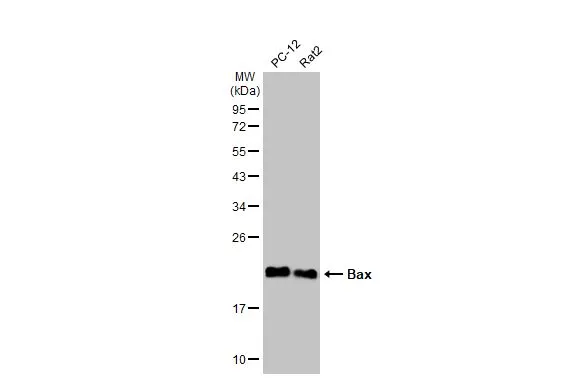
![Various tissue extracts (50 μg) were separated by 12% SDS-PAGE, and the membrane was blotted with Bax antibody [N1N2], N-term (GTX109683) diluted at 1:1000. The HRP-conjugated anti-rabbit IgG antibody (GTX213110-01) was used to detect the primary antibody.](https://www.genetex.com/upload/website/prouct_img/normal/GTX109683/GTX109683_44223_20220318_WB_M_R_w_23060500_920.webp "Various tissue extracts (50 μg) were separated by 12% SDS-PAGE, and the membrane was blotted with Bax antibody [N1N2], N-term (GTX109683) diluted at 1:1000. The HRP-conjugated anti-rabbit IgG antibody (GTX213110-01) was used to detect the primary antibody.")


Various whole cell extracts (30 μg) were separated by 12% SDS-PAGE, and the membrane was blotted with Bax antibody [N1N2], N-term (GTX109683) diluted at 1:1000. The HRP-conjugated anti-rabbit IgG antibody (GTX213110-01) was used to detect the primary antibody.
Bax antibody [N1N2], N-term
GTX109683
ApplicationsImmunoFluorescence, ImmunoPrecipitation, Western Blot, ImmunoCytoChemistry, ImmunoHistoChemistry, ImmunoHistoChemistry Frozen, ImmunoHistoChemistry Paraffin
Product group Antibodies
ReactivityHuman, Mouse, Rat
TargetBAX
Overview
- SupplierGeneTex
- Product NameBax antibody [N1N2], N-term
- Delivery Days Customer9
- Application Supplier NoteWB: 1:500-1:3000. ICC/IF: 1:100-1:1000. IHC-P: 1:100-1:1000. IP: 1:100-1:500. *Optimal dilutions/concentrations should be determined by the researcher.Not tested in other applications.
- ApplicationsImmunoFluorescence, ImmunoPrecipitation, Western Blot, ImmunoCytoChemistry, ImmunoHistoChemistry, ImmunoHistoChemistry Frozen, ImmunoHistoChemistry Paraffin
- CertificationResearch Use Only
- ClonalityPolyclonal
- Concentration1.27 mg/ml
- ConjugateUnconjugated
- Gene ID581
- Target nameBAX
- Target descriptionBCL2 associated X, apoptosis regulator
- Target synonymsBCL2L4, apoptosis regulator BAX, BCL2 associated X protein, BCL2-associated X protein omega, Baxdelta2(G8)-RFS protein, Baxdelta2G9, Baxdelta2G9omega, Baxdelta2omega, bcl-2-like protein 4, bcl2-L-4
- HostRabbit
- IsotypeIgG
- Protein IDQ07812
- Protein NameApoptosis regulator BAX
- Scientific DescriptionThe protein encoded by this gene belongs to the BCL2 protein family. BCL2 family members form hetero- or homodimers and act as anti- or pro-apoptotic regulators that are involved in a wide variety of cellular activities. This protein forms a heterodimer with BCL2, and functions as an apoptotic activator. This protein is reported to interact with, and increase the opening of, the mitochondrial voltage-dependent anion channel (VDAC), which leads to the loss in membrane potential and the release of cytochrome c. The expression of this gene is regulated by the tumor suppressor P53 and has been shown to be involved in P53-mediated apoptosis. Multiple alternatively spliced transcript variants, which encode different isoforms, have been reported for this gene. [provided by RefSeq]
- ReactivityHuman, Mouse, Rat
- Storage Instruction-20°C or -80°C,2°C to 8°C
- UNSPSC41116161
Datasheet
Related products
Product group Antibodies

Anti-Bax (active monomer) [6A7]Ab00120-1.1
ApplicationsImmunoPrecipitation, Western Blot, ImmunoHistoChemistry
ReactivityHuman, Mouse, Rat
TargetBAX
- SizePrice
Product group Antibodies
Anti-BAX AntibodyA101574
ApplicationsELISA, ImmunoHistoChemistry
ReactivityHuman
- SizePrice

Product group Antibodies
Anti-BAX AntibodyAMAB91490
ApplicationsWestern Blot, ImmunoHistoChemistry
ReactivityHuman
TargetBAX
- SizePrice

Product group Antibodies
Anti-Bax Antibody Picoband(r)A00183-CARRIER-FREE
ApplicationsFlow Cytometry, ImmunoFluorescence, Western Blot, ImmunoCytoChemistry, ImmunoHistoChemistry
ReactivityHuman, Mouse, Rat
TargetBAX
- SizePrice
Product group Antibodies
References
Bax Polyclonal AntibodyBS-0127R
ApplicationsFlow Cytometry, ImmunoFluorescence, ImmunoPrecipitation, Western Blot, ELISA, ImmunoCytoChemistry, ImmunoHistoChemistry, ImmunoHistoChemistry Frozen, ImmunoHistoChemistry Paraffin
ReactivityBovine, Canine, Human, Mouse, Porcine, Rabbit, Rat, Sheep
TargetBAX
- SizePrice
Product group Antibodies
BAX Monoclonal AntibodyCSB-MA183751
ApplicationsWestern Blot, ELISA, ImmunoHistoChemistry
ReactivityHuman, Mouse, Rat
TargetBAX
- SizePrice
Product group Antibodies
ApplicationsImmunoPrecipitation, Western Blot, ImmunoCytoChemistry, ImmunoHistoChemistry
ReactivityMouse, Porcine, Rat
TargetBAX
- SizePrice
Product group Antibodies
Bax antibody [2D3]GTX16119
ApplicationsWestern Blot
ReactivityBovine, Canine, Feline, Human, Rat
TargetBAX
- SizePrice
Product group Antibodies
Bax antibodyGTX16143
ApplicationsImmunoPrecipitation
ReactivityHuman
TargetBAX
- SizePrice