


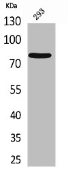
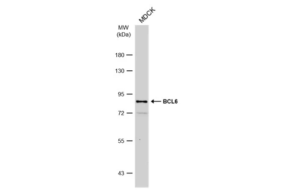
Figure 1. Western blot analysis of Bcl6 using anti-Bcl6 antibody (A00142-1). Electrophoresis was performed on a 5-20% SDS-PAGE gel at 70V (Stacking gel) / 90V (Resolving gel) for 2-3 hours. The sample well of each lane was loaded with 50ug of sample under reducing conditions. Lane 1: human Raji whole cell lysate, Lane 2: human MCF-7 whole cell lysate, Lane 3: human placenta tissue lysate, Lane 4: human A549 whole cell lysate, Lane 5: human Caco-2 whole cell lysate, Lane 6: human U2OS whole cell lysate. After Electrophoresis, proteins were transferred to a Nitrocellulose membrane at 150mA for 50-90 minutes. Blocked the membrane with 5% Non-fat Milk/ TBS for 1.5 hour at RT. The membrane was incubated with rabbit anti-Bcl6 antigen affinity purified polyclonal antibody (Catalog # A00142-1) at 0.5 microg/mL overnight at 4°C, then washed with TBS-0.1%Tween 3 times with 5 minutes each and probed with a goat anti-rabbit IgG-HRP secondary antibody at a dilution of 1:10000 for 1.5 hour at RT. The signal is developed using an Enhanced Chemiluminescent detection (ECL) kit (Catalog # EK1002) with Tanon 5200 system. A specific band was detected for Bcl6 at approximately 88KD. The expected band size for Bcl6 is at 79KD.
. Electrophoresis was performed on a 5-20% SDS-PAGE gel at 70V (Stacking gel) / 90V (Resolving gel) for 2-3 hours. The sample well of each lane was loaded with 50ug of sample under reducing conditions. Lane 1: rat testis tissue lysate, Lane 2: rat spleen tissue lysate, Lane 3: rat brain tissue lysate, Lane 4: mouse testis tissue lysate, Lane 5: mouse spleen tissue lysate, Lane 6: mouse brain tissue lysate. After Electrophoresis, proteins were transferred to a Nitrocellulose membrane at 150mA for 50-90 minutes. Blocked the membrane with 5% Non-fat Milk/ TBS for 1.5 hour at RT. The membrane was incubated with rabbit anti-Bcl6 antigen affinity purified polyclonal antibody (Catalog # A00142-1) at 0.5 microg/mL overnight at 4°C, then washed with TBS-0.1%Tween 3 times with 5 minutes each and probed with a goat anti-rabbit IgG-HRP secondary antibody at a dilution of 1:10000 for 1.5 hour at RT. The signal is developed using an Enhanced Chemiluminescent detection (ECL) kit (Catalog # EK1002) with Tanon 5200 system. A specific band was detected for Bcl6 at approximately 88KD. The expected band size for Bcl6 is at 79KD.")
. Bcl6 was detected in paraffin-embedded section of human tonsil tissues. Heat mediated antigen retrieval was performed in citrate buffer (pH6, epitope retrieval solution) for 20 mins. The tissue section was blocked with 10% goat serum. The tissue section was then incubated with 1microg/ml rabbit anti-Bcl6 Antibody (A00142-1) overnight at 4°C. Biotinylated goat anti-rabbit IgG was used as secondary antibody and incubated for 30 minutes at 37°C. The tissue section was developed using Strepavidin-Biotin-Complex (SABC)(Catalog # SA1022) with DAB as the chromogen.")
. Bcl6 was detected in paraffin-embedded section of mouse spleen tissues. Heat mediated antigen retrieval was performed in citrate buffer (pH6, epitope retrieval solution) for 20 mins. The tissue section was blocked with 10% goat serum. The tissue section was then incubated with 1microg/ml rabbit anti-Bcl6 Antibody (A00142-1) overnight at 4°C. Biotinylated goat anti-rabbit IgG was used as secondary antibody and incubated for 30 minutes at 37°C. The tissue section was developed using Strepavidin-Biotin-Complex (SABC)(Catalog # SA1022) with DAB as the chromogen.")
. Overlay histogram showing BRL cells stained with A00142-1 (Blue line). To facilitate intracellular staining, cells were fixed with 4% paraformaldehyde and permeabilized with permeabilization buffer. The cells were blocked with 10% normal goat serum. And then incubated with rabbit anti-Bcl6 Antibody (A00142-1,1microg/1x106 cells) for 30 min at 20°C. DyLight®488 conjugated goat anti-rabbit IgG (BA1127, 5-10microg/1x106 cells) was used as secondary antibody for 30 minutes at 20°C. Isotype control antibody (Green line) was rabbit IgG (1microg/1x106) used under the same conditions. Unlabelled sample (Red line) was also used as a control.")
Figure 1. Western blot analysis of Bcl6 using anti-Bcl6 antibody (A00142-1). Electrophoresis was performed on a 5-20% SDS-PAGE gel at 70V (Stacking gel) / 90V (Resolving gel) for 2-3 hours. The sample well of each lane was loaded with 50ug of sample under reducing conditions. Lane 1: human Raji whole cell lysate, Lane 2: human MCF-7 whole cell lysate, Lane 3: human placenta tissue lysate, Lane 4: human A549 whole cell lysate, Lane 5: human Caco-2 whole cell lysate, Lane 6: human U2OS whole cell lysate. After Electrophoresis, proteins were transferred to a Nitrocellulose membrane at 150mA for 50-90 minutes. Blocked the membrane with 5% Non-fat Milk/ TBS for 1.5 hour at RT. The membrane was incubated with rabbit anti-Bcl6 antigen affinity purified polyclonal antibody (Catalog # A00142-1) at 0.5 microg/mL overnight at 4°C, then washed with TBS-0.1%Tween 3 times with 5 minutes each and probed with a goat anti-rabbit IgG-HRP secondary antibody at a dilution of 1:10000 for 1.5 hour at RT. The signal is developed using an Enhanced Chemiluminescent detection (ECL) kit (Catalog # EK1002) with Tanon 5200 system. A specific band was detected for Bcl6 at approximately 88KD. The expected band size for Bcl6 is at 79KD.
Anti-BCL6 Antibody Picoband(r)
A00142-1-CARRIER-FREE
ApplicationsFlow Cytometry, Western Blot, ImmunoCytoChemistry, ImmunoHistoChemistry
Product group Antibodies
ReactivityHuman, Mouse, Rat
TargetBCL6
Overview
- SupplierBoster Bio
- Product NameAnti-BCL6 Antibody Picoband(r)
- Delivery Days Customer9
- ApplicationsFlow Cytometry, Western Blot, ImmunoCytoChemistry, ImmunoHistoChemistry
- CertificationResearch Use Only
- ClonalityPolyclonal
- Concentration500 ug/ml
- Gene ID604
- Target nameBCL6
- Target descriptionBCL6 transcription repressor
- Target synonymsBCL5, BCL6A, LAZ3, ZBTB27, ZNF51, B-cell lymphoma 6 protein, B cell CLL/lymphoma 6, B-cell lymphoma 5 protein, B-cell lymphoma 6 protein transcript, BCL-5, BCL-6, cys-his2 zinc finger transcription factor, lymphoma-associated zinc finger gene on chromosome 3, protein LAZ-3, zinc finger and BTB domain-containing protein 27, zinc finger protein 51, zinc finger transcription factor BCL6S
- HostRabbit
- IsotypeIgG
- Protein IDP41182
- Protein NameB-cell lymphoma 6 protein
- Scientific DescriptionBoster Bio Anti-BCL6 Antibody Picoband® catalog # A00142-1. Tested in Flow Cytometry, IHC, ICC, WB applications. This antibody reacts with Human, Mouse, Rat. The brand Picoband indicates this is a premium antibody that guarantees superior quality, high affinity, and strong signals with minimal background in Western blot applications. Only our best-performing antibodies are designated as Picoband, ensuring unmatched performance.
- ReactivityHuman, Mouse, Rat
- Storage Instruction-20°C,2°C to 8°C
- UNSPSC12352203
Related products
Product group Antibodies
Anti-Bcl-6 AntibodyA97690
ApplicationsWestern Blot, ELISA
ReactivityHuman, Mouse, Rat
- SizePrice

Product group Antibodies
Anti-BCL6 Antibody144-66311
ApplicationsImmunoFluorescence, Western Blot
ReactivityHuman, Mouse
TargetBCL6
- SizePrice
Product group Antibodies
Anti-BCL6 AntibodyAMAB91977
ApplicationsImmunoCytoChemistry, ImmunoHistoChemistry
ReactivityHuman
TargetBCL6
- SizePrice

Product group Antibodies
References
Bcl-6 Polyclonal AntibodyBS-2734R
ApplicationsImmunoFluorescence, Western Blot, ELISA, ImmunoHistoChemistry, ImmunoHistoChemistry Frozen, ImmunoHistoChemistry Paraffin
ReactivityHuman, Mouse
TargetBCL6
- SizePrice
Product group Antibodies
BCL6 AntibodyCSB-PA005439
ApplicationsWestern Blot, ELISA
ReactivityHuman, Mouse, Rat
TargetBCL6
- SizePrice
Product group Antibodies
ApplicationsImmunoPrecipitation, Western Blot, ImmunoCytoChemistry, ImmunoHistoChemistry
TargetBCL6
- SizePrice
Product group Antibodies
BCL6 antibody [N2C1], InternalGTX101338
ApplicationsImmunoPrecipitation, Western Blot, ChIP Chromatin ImmunoPrecipitation, ImmunoHistoChemistry, ImmunoHistoChemistry Paraffin
ReactivityCanine, Human, Mouse, Rat
TargetBCL6
- SizePrice
Product group Antibodies

BCL6 Antibody (Internal)LS-C368565
ApplicationsWestern Blot, ImmunoHistoChemistry
ReactivityCanine, Human, Mouse, Rat
TargetBCL6
- SizePrice