


Figure 1. Western blot analysis of BPIFB1 using anti-BPIFB1 antibody (A08872-1). Electrophoresis was performed on a 5-20% SDS-PAGE gel at 70V (Stacking gel) / 90V (Resolving gel) for 2-3 hours. The sample well of each lane was loaded with 30 ug of sample under reducing conditions. Lane 1: human MCF-7 whole cell lysates, Lane 2: human HepG2 whole cell lysates, Lane 3: human A549 whole cell lysates. After electrophoresis, proteins were transferred to a nitrocellulose membrane at 150 mA for 50-90 minutes. Blocked the membrane with 5% non-fat milk/TBS for 1.5 hour at RT. The membrane was incubated with rabbit anti-BPIFB1 antigen affinity purified polyclonal antibody (Catalog # A08872-1) at 0.5 microg/mL overnight at 4°C, then washed with TBS-0.1%Tween 3 times with 5 minutes each and probed with a goat anti-rabbit IgG-HRP secondary antibody at a dilution of 1:5000 for 1.5 hour at RT. The signal is developed using an Enhanced Chemiluminescent detection (ECL) kit (Catalog # EK1002) with Tanon 5200 system. A specific band was detected for BPIFB1 at approximately 52 kDa. The expected band size for BPIFB1 is at 52 kDa.
. Overlay histogram showing HepG2 cells stained with A08872-1 (Blue line). The cells were fixed with 4% paraformaldehyde and blocked with 10% normal goat serum. And then incubated with rabbit anti-BPIFB1 Antibody (A08872-1, 1 microg/1x106 cells) for 30 min at 20°C. DyLight®488 conjugated goat anti-rabbit IgG (BA1127, 5-10 microg/1x106 cells) was used as secondary antibody for 30 minutes at 20°C. Isotype control antibody (Green line) was rabbit IgG (1 microg/1x106) used under the same conditions. Unlabelled sample (Red line) was also used as a control.")
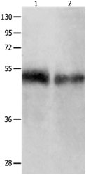
Figure 1. Western blot analysis of BPIFB1 using anti-BPIFB1 antibody (A08872-1). Electrophoresis was performed on a 5-20% SDS-PAGE gel at 70V (Stacking gel) / 90V (Resolving gel) for 2-3 hours. The sample well of each lane was loaded with 30 ug of sample under reducing conditions. Lane 1: human MCF-7 whole cell lysates, Lane 2: human HepG2 whole cell lysates, Lane 3: human A549 whole cell lysates. After electrophoresis, proteins were transferred to a nitrocellulose membrane at 150 mA for 50-90 minutes. Blocked the membrane with 5% non-fat milk/TBS for 1.5 hour at RT. The membrane was incubated with rabbit anti-BPIFB1 antigen affinity purified polyclonal antibody (Catalog # A08872-1) at 0.5 microg/mL overnight at 4°C, then washed with TBS-0.1%Tween 3 times with 5 minutes each and probed with a goat anti-rabbit IgG-HRP secondary antibody at a dilution of 1:5000 for 1.5 hour at RT. The signal is developed using an Enhanced Chemiluminescent detection (ECL) kit (Catalog # EK1002) with Tanon 5200 system. A specific band was detected for BPIFB1 at approximately 52 kDa. The expected band size for BPIFB1 is at 52 kDa.
Anti-BPIFB1 Antibody Picoband(r)
A08872-1-CARRIER-FREE
ApplicationsFlow Cytometry, Western Blot, ELISA
Product group Antibodies
ReactivityHuman
TargetBPIFB1
Overview
- SupplierBoster Bio
- Product NameAnti-BPIFB1 Antibody Picoband(r)
- Delivery Days Customer9
- ApplicationsFlow Cytometry, Western Blot, ELISA
- CertificationResearch Use Only
- ClonalityPolyclonal
- Concentration500 ug/ml
- Gene ID92747
- Target nameBPIFB1
- Target descriptionBPI fold containing family B member 1
- Target synonymsC20orf114, LPLUNC1, BPI fold-containing family B member 1, VEMSGP, long palate, lung and nasal epithelium carcinoma associated 1, long palate, lung and nasal epithelium carcinoma-associated protein 1, von Ebner minor salivary gland protein
- HostRabbit
- Protein IDQ8TDL5
- Protein NameBPI fold-containing family B member 1
- Scientific DescriptionBoster Bio Anti-BPIFB1 Antibody Picoband® catalog # A08872-1. Tested in WB, Flow Cytometry, ELISA applications. This antibody reacts with Human. The brand Picoband indicates this is a premium antibody that guarantees superior quality, high affinity, and strong signals with minimal background in Western blot applications. Only our best-performing antibodies are designated as Picoband, ensuring unmatched performance.
- ReactivityHuman
- Storage Instruction-20°C,2°C to 8°C
- UNSPSC12352203
Related products
Product group Antibodies
Anti-BPIFB1 AntibodyA47686
ApplicationsWestern Blot, ELISA, ImmunoHistoChemistry
ReactivityHuman
- SizePrice
Product group Antibodies
LPLUNC1 Polyclonal AntibodyBS-0976R
ApplicationsImmunoFluorescence, Western Blot, ELISA, ImmunoCytoChemistry, ImmunoHistoChemistry, ImmunoHistoChemistry Frozen, ImmunoHistoChemistry Paraffin
ReactivityHuman, Mouse, Rat
TargetBPIFB1
- SizePrice
Product group Antibodies
BPIFB1 AntibodyCSB-PA072508
ApplicationsELISA, ImmunoHistoChemistry
ReactivityHuman
TargetBPIFB1
- SizePrice

Product group Antibodies
Anti-BPIFB1 AntibodyHPA024256
ApplicationsImmunoHistoChemistry
ReactivityHuman
TargetBPIFB1
- SizePrice
![FACS analysis of HeLa cells using GTX60533 LPlunc1 antibody [2A5]. Green : LPlunc1 Red : negative control](https://www.genetex.com/upload/website/prouct_img/normal/GTX60533/GTX60533_20170912_FACS_w_23061123_673.webp)
Product group Antibodies
Lplunc1 antibody [2A5]GTX60533
ApplicationsFlow Cytometry, ImmunoFluorescence, Western Blot, ELISA, ImmunoCytoChemistry, ImmunoHistoChemistry, ImmunoHistoChemistry Paraffin
ReactivityHuman
TargetBPIFB1
- SizePrice
Product group Antibodies

BPIFB1 Antibody (Preservative Free)LS-C817789
ApplicationsELISA, ImmunoHistoChemistry, ImmunoHistoChemistry Paraffin
ReactivityHuman
TargetBPIFB1
- SizePrice