

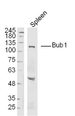
Anti-BUB1 Antibody
A101535
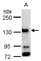
ApplicationsWestern Blot, ELISA
Product group Antibodies
ReactivityHuman
Overview
- SupplierAntibodies.com
- Product NameAnti-BUB1 Antibody
- Delivery Days Customer7
- ApplicationsWestern Blot, ELISA
- CertificationResearch Use Only
- ClonalityPolyclonal
- ConjugateUnconjugated
- HostRabbit
- IsotypeIgG
- Scientific DescriptionRabbit polyclonal antibody to BUB1.
- ReactivityHuman
- UNSPSC12352203
Related products

Product group Antibodies
Anti-BUB1 Antibody Picoband(r)A00776-3-CARRIER-FREE
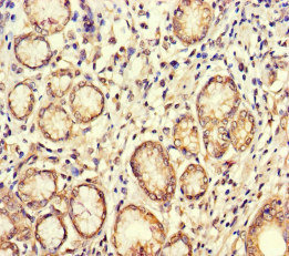
ApplicationsWestern Blot, ELISA, ImmunoHistoChemistry
ReactivityHuman, Mouse, Rat
TargetBUB1
- SizePrice
Product group Antibodies
Anti-BUB1 Antibody144-01929
ApplicationsWestern Blot, ImmunoHistoChemistry
ReactivityHuman, Mouse, Rat
TargetBUB1
- SizePrice
Product group Antibodies
Bub1 Polyclonal AntibodyBS-4294R
ApplicationsImmunoFluorescence, Western Blot, ELISA, ImmunoCytoChemistry, ImmunoHistoChemistry, ImmunoHistoChemistry Frozen, ImmunoHistoChemistry Paraffin
ReactivityBovine, Canine, Equine, Human, Mouse, Rabbit, Rat
TargetBUB1
- SizePrice
Product group Antibodies
BUB1 AntibodyCSB-PA002882LA01HU
ApplicationsELISA, ImmunoHistoChemistry
ReactivityHuman
TargetBUB1
- SizePrice
Product group Antibodies
Bub1 Recombinant AntibodyCAC12031
ApplicationsWestern Blot, ELISA
TargetBUB1
- SizePrice
Product group Antibodies

BUB1 AntibodyLS-C401461
ApplicationsELISA, ImmunoHistoChemistry
ReactivityHuman
TargetBUB1
- SizePrice
Product group Antibodies
BUB1 antibodyGTX107497
ApplicationsImmunoFluorescence, Western Blot, ImmunoCytoChemistry
ReactivityHuman
TargetBUB1
- SizePrice
Product group Antibodies
ApplicationsImmunoFluorescence, Western Blot, ELISA, ImmunoCytoChemistry, ImmunoHistoChemistry, ImmunoHistoChemistry Paraffin
ReactivityHuman
TargetBUB1
- SizePrice