


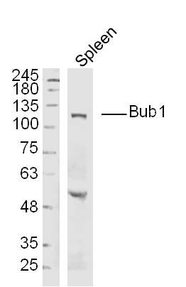
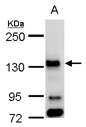
Figure 1. Western blot analysis of BUB1 using anti-BUB1 antibody (A00776-3). Electrophoresis was performed on a 5-20% SDS-PAGE gel at 70V (Stacking gel) / 90V (Resolving gel) for 2-3 hours. The sample well of each lane was loaded with 30 ug of sample under reducing conditions. Lane 1: human Hela whole cell lysates, Lane 2: human 293T whole cell lysates, Lane 3: human K562 whole cell lysates, Lane 4: mouse testis tissue lysates. After electrophoresis, proteins were transferred to a nitrocellulose membrane at 150 mA for 50-90 minutes. Blocked the membrane with 5% non-fat milk/TBS for 1.5 hour at RT. The membrane was incubated with rabbit anti-BUB1 antigen affinity purified polyclonal antibody (Catalog # A00776-3) at 0.5 microg/mL overnight at 4°C, then washed with TBS-0.1%Tween 3 times with 5 minutes each and probed with a goat anti-rabbit IgG-HRP secondary antibody at a dilution of 1:5000 for 1.5 hour at RT. The signal is developed using an Enhanced Chemiluminescent detection (ECL) kit (Catalog # EK1002) with Tanon 5200 system. A specific band was detected for BUB1 at approximately 130 kDa. The expected band size for BUB1 is at 122 kDa.
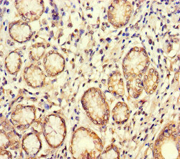
. BUB1 was detected in a paraffin-embedded section of human bladder cancer tissue. Heat mediated antigen retrieval was performed in EDTA buffer (pH 8.0, epitope retrieval solution). The tissue section was blocked with 10% goat serum. The tissue section was then incubated with 2 microg/ml rabbit anti-BUB1 Antibody (A00776-3) overnight at 4°C. Biotinylated goat anti-rabbit IgG was used as secondary antibody and incubated for 30 minutes at 37°C. The tissue section was developed using Strepavidin-Biotin-Complex (SABC) (Catalog # SA1022) with DAB as the chromogen.")
. BUB1 was detected in a paraffin-embedded section of mouse thymus tissue. Heat mediated antigen retrieval was performed in EDTA buffer (pH 8.0, epitope retrieval solution). The tissue section was blocked with 10% goat serum. The tissue section was then incubated with 2 microg/ml rabbit anti-BUB1 Antibody (A00776-3) overnight at 4°C. Biotinylated goat anti-rabbit IgG was used as secondary antibody and incubated for 30 minutes at 37°C. The tissue section was developed using Strepavidin-Biotin-Complex (SABC) (Catalog # SA1022) with DAB as the chromogen.")
. BUB1 was detected in a paraffin-embedded section of rat thymus tissue. Heat mediated antigen retrieval was performed in EDTA buffer (pH 8.0, epitope retrieval solution). The tissue section was blocked with 10% goat serum. The tissue section was then incubated with 2 microg/ml rabbit anti-BUB1 Antibody (A00776-3) overnight at 4°C. Biotinylated goat anti-rabbit IgG was used as secondary antibody and incubated for 30 minutes at 37°C. The tissue section was developed using Strepavidin-Biotin-Complex (SABC) (Catalog # SA1022) with DAB as the chromogen.")
Figure 1. Western blot analysis of BUB1 using anti-BUB1 antibody (A00776-3). Electrophoresis was performed on a 5-20% SDS-PAGE gel at 70V (Stacking gel) / 90V (Resolving gel) for 2-3 hours. The sample well of each lane was loaded with 30 ug of sample under reducing conditions. Lane 1: human Hela whole cell lysates, Lane 2: human 293T whole cell lysates, Lane 3: human K562 whole cell lysates, Lane 4: mouse testis tissue lysates. After electrophoresis, proteins were transferred to a nitrocellulose membrane at 150 mA for 50-90 minutes. Blocked the membrane with 5% non-fat milk/TBS for 1.5 hour at RT. The membrane was incubated with rabbit anti-BUB1 antigen affinity purified polyclonal antibody (Catalog # A00776-3) at 0.5 microg/mL overnight at 4°C, then washed with TBS-0.1%Tween 3 times with 5 minutes each and probed with a goat anti-rabbit IgG-HRP secondary antibody at a dilution of 1:5000 for 1.5 hour at RT. The signal is developed using an Enhanced Chemiluminescent detection (ECL) kit (Catalog # EK1002) with Tanon 5200 system. A specific band was detected for BUB1 at approximately 130 kDa. The expected band size for BUB1 is at 122 kDa.
Anti-BUB1 Antibody Picoband(r)
A00776-3-CARRIER-FREE
ApplicationsWestern Blot, ELISA, ImmunoHistoChemistry
Product group Antibodies
ReactivityHuman, Mouse, Rat
TargetBUB1
Overview
- SupplierBoster Bio
- Product NameAnti-BUB1 Antibody Picoband(r)
- Delivery Days Customer9
- ApplicationsWestern Blot, ELISA, ImmunoHistoChemistry
- CertificationResearch Use Only
- ClonalityPolyclonal
- Concentration500 ug/ml
- Gene ID699
- Target nameBUB1
- Target descriptionBUB1 mitotic checkpoint serine/threonine kinase
- Target synonymsBUB1A, BUB1L, MCPH30, hBUB1, mitotic checkpoint serine/threonine-protein kinase BUB1, BUB1 budding uninhibited by benzimidazoles 1 homolog, budding uninhibited by benzimidazoles 1 homolog, mitotic spindle checkpoint kinase, putative serine/threonine-protein kinase
- HostRabbit
- IsotypeIgG
- Protein IDO43683
- Protein NameMitotic checkpoint serine/threonine-protein kinase BUB1
- Scientific DescriptionBoster Bio Anti-BUB1 Antibody Picoband® catalog # A00776-3. Tested in ELISA, IHC, WB applications. This antibody reacts with Human, Mouse, Rat. The brand Picoband indicates this is a premium antibody that guarantees superior quality, high affinity, and strong signals with minimal background in Western blot applications. Only our best-performing antibodies are designated as Picoband, ensuring unmatched performance.
- ReactivityHuman, Mouse, Rat
- Storage Instruction-20°C,2°C to 8°C
- UNSPSC12352203
Related products
Product group Antibodies
Anti-BUB1 AntibodyA101535
ApplicationsWestern Blot, ELISA
ReactivityHuman
- SizePrice
Product group Antibodies
Anti-BUB1 Antibody144-01929
ApplicationsWestern Blot, ImmunoHistoChemistry
ReactivityHuman, Mouse, Rat
TargetBUB1
- SizePrice
Product group Antibodies
Bub1 Polyclonal AntibodyBS-4294R
ApplicationsImmunoFluorescence, Western Blot, ELISA, ImmunoCytoChemistry, ImmunoHistoChemistry, ImmunoHistoChemistry Frozen, ImmunoHistoChemistry Paraffin
ReactivityBovine, Canine, Equine, Human, Mouse, Rabbit, Rat
TargetBUB1
- SizePrice
Product group Antibodies
BUB1 AntibodyCSB-PA002882LA01HU
ApplicationsELISA, ImmunoHistoChemistry
ReactivityHuman
TargetBUB1
- SizePrice
Product group Antibodies
Bub1 Recombinant AntibodyCAC12031
ApplicationsWestern Blot, ELISA
TargetBUB1
- SizePrice
Product group Antibodies

BUB1 AntibodyLS-C401461
ApplicationsELISA, ImmunoHistoChemistry
ReactivityHuman
TargetBUB1
- SizePrice
Product group Antibodies
BUB1 antibodyGTX107497
ApplicationsImmunoFluorescence, Western Blot, ImmunoCytoChemistry
ReactivityHuman
TargetBUB1
- SizePrice
Product group Antibodies
ApplicationsImmunoFluorescence, Western Blot, ELISA, ImmunoCytoChemistry, ImmunoHistoChemistry, ImmunoHistoChemistry Paraffin
ReactivityHuman
TargetBUB1
- SizePrice