


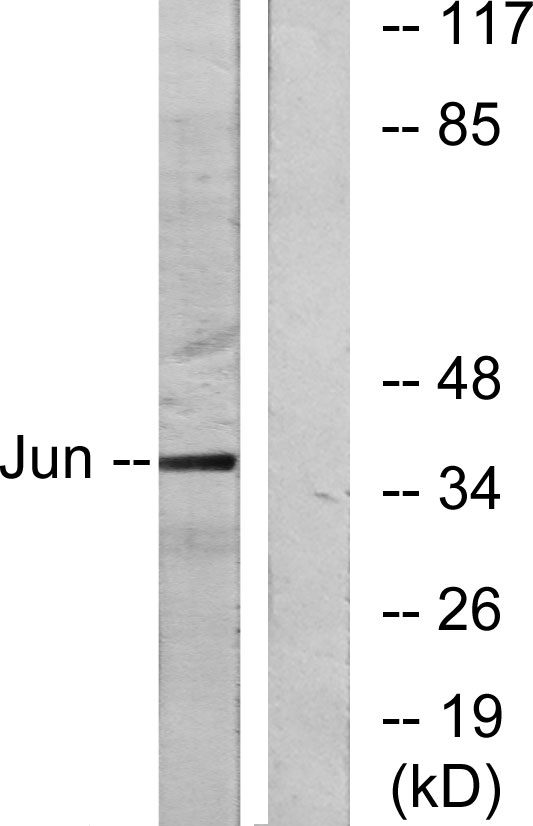
Figure 1. Western blot analysis of C-Jun/JUN using anti-C-Jun/JUN antibody (A02038-3). Electrophoresis was performed on a 5-20% SDS-PAGE gel at 70V (Stacking gel) / 90V (Resolving gel) for 2-3 hours. The sample well of each lane was loaded with 30 ug of sample under reducing conditions. Lane 1: human Hela whole cell lysates, Lane 2: human K562 whole cell lysates, Lane 3: human Jurkat whole cell lysates, Lane 4: human U-87MG whole cell lysates. After electrophoresis, proteins were transferred to a nitrocellulose membrane at 150 mA for 50-90 minutes. Blocked the membrane with 5% non-fat milk/TBS for 1.5 hour at RT. The membrane was incubated with rabbit anti-C-Jun/JUN antigen affinity purified polyclonal antibody (Catalog # A02038-3) at 0.5 microg/mL overnight at 4°C, then washed with TBS-0.1%Tween 3 times with 5 minutes each and probed with a goat anti-rabbit IgG-HRP secondary antibody at a dilution of 1:5000 for 1.5 hour at RT. The signal is developed using an Enhanced Chemiluminescent detection (ECL) kit (Catalog # EK1002) with Tanon 5200 system. A specific band was detected for C-Jun/JUN at approximately 36 kDa. The expected band size for C-Jun/JUN is at 36 kDa.
. C-Jun/JUN was detected in a paraffin-embedded section of human renal cell carcinoma tissue. Heat mediated antigen retrieval was performed in EDTA buffer (pH 8.0, epitope retrieval solution). The tissue section was blocked with 10% goat serum. The tissue section was then incubated with 2 microg/ml rabbit anti-C-Jun/JUN Antibody (A02038-3) overnight at 4°C. Peroxidase Conjugated Goat Anti-rabbit IgG was used as secondary antibody and incubated for 30 minutes at 37°C. The tissue section was developed using HRP Conjugated Rabbit IgG Super Vision Assay Kit (Catalog # SV0002) with DAB as the chromogen.")
. C-Jun/JUN was detected in a paraffin-embedded section of human the renal pelvis squamous metaplasia tissue. Heat mediated antigen retrieval was performed in EDTA buffer (pH 8.0, epitope retrieval solution). The tissue section was blocked with 10% goat serum. The tissue section was then incubated with 2 microg/ml rabbit anti-C-Jun/JUN Antibody (A02038-3) overnight at 4°C. Peroxidase Conjugated Goat Anti-rabbit IgG was used as secondary antibody and incubated for 30 minutes at 37°C. The tissue section was developed using HRP Conjugated Rabbit IgG Super Vision Assay Kit (Catalog # SV0002) with DAB as the chromogen.")
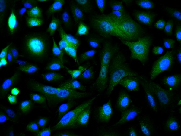
 and anti-Beta Tubulin antibody (M01857-3). C-Jun/JUN was detected in immunocytochemical section of A431 cells. Enzyme antigen retrieval was performed using IHC enzyme antigen retrieval reagent (AR0022) for 15 mins. The cells were blocked with 10% goat serum. And then incubated with 5 microg/mL rabbit anti-C-Jun/JUN beta Antibody (A02038-3) and mouse anti-Beta Tubulin antibody (M01857-3) overnight at 4°C. DyLight®488 Conjugated Goat Anti-Rabbit IgG (BA1127) and DyLight®594 Conjugated Goat Anti-Mouse IgG (BA1141) were used as secondary antibody at 1:100 dilution and incubated for 30 minutes at 37°C. Visualize using a fluorescence microscope and filter sets appropriate for the label used.")
. Overlay histogram showing U20S cells stained with A02038-3 (Blue line). To facilitate intracellular staining, cells were fixed with 4% paraformaldehyde and permeabilized with permeabilization buffer. The cells were blocked with 10% normal goat serum. And then incubated with rabbit anti-C-Jun/JUN Antibody (A02038-3, 1 microg/1x106 cells) for 30 min at 20°C. DyLight®488 conjugated goat anti-rabbit IgG (BA1127, 5-10 microg/1x106 cells) was used as secondary antibody for 30 minutes at 20°C. Isotype control antibody (Green line) was rabbit IgG (1 microg/1x106) used under the same conditions. Unlabelled sample without incubation with primary antibody and secondary antibody (Red line) was used as a blank control.")
Figure 1. Western blot analysis of C-Jun/JUN using anti-C-Jun/JUN antibody (A02038-3). Electrophoresis was performed on a 5-20% SDS-PAGE gel at 70V (Stacking gel) / 90V (Resolving gel) for 2-3 hours. The sample well of each lane was loaded with 30 ug of sample under reducing conditions. Lane 1: human Hela whole cell lysates, Lane 2: human K562 whole cell lysates, Lane 3: human Jurkat whole cell lysates, Lane 4: human U-87MG whole cell lysates. After electrophoresis, proteins were transferred to a nitrocellulose membrane at 150 mA for 50-90 minutes. Blocked the membrane with 5% non-fat milk/TBS for 1.5 hour at RT. The membrane was incubated with rabbit anti-C-Jun/JUN antigen affinity purified polyclonal antibody (Catalog # A02038-3) at 0.5 microg/mL overnight at 4°C, then washed with TBS-0.1%Tween 3 times with 5 minutes each and probed with a goat anti-rabbit IgG-HRP secondary antibody at a dilution of 1:5000 for 1.5 hour at RT. The signal is developed using an Enhanced Chemiluminescent detection (ECL) kit (Catalog # EK1002) with Tanon 5200 system. A specific band was detected for C-Jun/JUN at approximately 36 kDa. The expected band size for C-Jun/JUN is at 36 kDa.
Anti-c-Jun/JUN Antibody Picoband(r)
A02038-3-CY3
ApplicationsFlow Cytometry, ImmunoFluorescence, Western Blot, ELISA, ImmunoCytoChemistry, ImmunoHistoChemistry
Product group Antibodies
ReactivityHuman
TargetJUN
Overview
- SupplierBoster Bio
- Product NameAnti-c-Jun/JUN Antibody Picoband(r)
- Delivery Days Customer9
- ApplicationsFlow Cytometry, ImmunoFluorescence, Western Blot, ELISA, ImmunoCytoChemistry, ImmunoHistoChemistry
- CertificationResearch Use Only
- ClonalityPolyclonal
- Concentration500 ug/ml
- ConjugateCy3
- Gene ID3725
- Target nameJUN
- Target descriptionJun proto-oncogene, AP-1 transcription factor subunit
- Target synonymsAP-1, AP1, c-Jun, cJUN, p39, transcription factor Jun, Jun activation domain binding protein, activator protein 1, enhancer-binding protein AP1, jun oncogene, proto-oncogene c-Jun, proto-oncogene cJun, transcription factor AP-1, transcription factor AP-1 subunit Jun, v-jun avian sarcoma virus 17 oncogene homolog, v-jun sarcoma virus 17 oncogene homolog
- HostRabbit
- IsotypeIgG
- Protein IDP05412
- Protein NameTranscription factor Jun
- Scientific DescriptionBoster Bio Anti-c-Jun/JUN Antibody Picoband® catalog # A02038-3. Tested in ELISA, Flow Cytometry, IF, IHC, ICC, WB applications. This antibody reacts with Human. The brand Picoband indicates this is a premium antibody that guarantees superior quality, high affinity, and strong signals with minimal background in Western blot applications. Only our best-performing antibodies are designated as Picoband, ensuring unmatched performance.
- ReactivityHuman
- Storage Instruction-20°C,2°C to 8°C
- UNSPSC12352203
Related products
Product group Antibodies
Jun Polyclonal AntibodyCAC09196
ApplicationsImmunoFluorescence, Western Blot, ELISA
ReactivityMouse, Rat
TargetJUN
- SizePrice
Product group Antibodies
References
c-Jun Polyclonal AntibodyBS-0670R
ApplicationsFlow Cytometry, ImmunoFluorescence, Western Blot, ELISA, ImmunoCytoChemistry, ImmunoHistoChemistry, ImmunoHistoChemistry Frozen, ImmunoHistoChemistry Paraffin
ReactivityBovine, Canine, Chicken, Human, Mouse, Porcine, Rat, Sheep
TargetJUN
- SizePrice
Product group Antibodies
Anti-c-Jun Antibody144-61581
ApplicationsImmunoFluorescence, Western Blot
ReactivityHuman, Mouse, Rat
TargetJUN
- SizePrice

Product group Antibodies
Anti-JUN AntibodyAMAB91587
ApplicationsImmunoCytoChemistry, ImmunoHistoChemistry
ReactivityHuman
TargetJUN
- SizePrice
Product group Antibodies
Anti-c-Jun AntibodyA95242
ApplicationsImmunoPrecipitation, Western Blot, ELISA, ImmunoHistoChemistry
ReactivityHuman, Mouse, Rat
- SizePrice
Product group Antibodies
References
c-Jun antibodyGTX112974
ApplicationsImmunoFluorescence, Western Blot, ImmunoCytoChemistry, ImmunoHistoChemistry, ImmunoHistoChemistry Paraffin
ReactivityHuman
TargetJUN
- SizePrice
Product group Antibodies

JUN / c-Jun Antibody (C-Terminus)LS-C368398
ApplicationsWestern Blot, ImmunoHistoChemistry
ReactivityBovine, Chicken, Human, Mouse, Porcine, Rat
TargetJUN
- SizePrice