


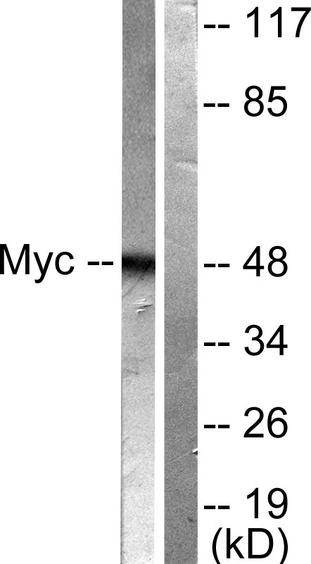
Figure 1. Western blot analysis of C-Myc/MYC using anti-C-Myc/MYC antibody (A00026-1). Electrophoresis was performed on a 5-20% SDS-PAGE gel at 70V (Stacking gel) / 90V (Resolving gel) for 2-3 hours. The sample well of each lane was loaded with 30 ug of sample under reducing conditions. Lane 1: human Raji whole cell lysates, Lane 2: human K562 whole cell lysates, Lane 3: rat brain tissue lysates, Lane 4: rat C6 whole cell lysates, Lane 5: mouse brain tissue lysates, Lane 6: mouse NIH/3T3 whole cell lysates. After electrophoresis, proteins were transferred to a nitrocellulose membrane at 150 mA for 50-90 minutes. Blocked the membrane with 5% non-fat milk/TBS for 1.5 hour at RT. The membrane was incubated with rabbit anti-C-Myc/MYC antigen affinity purified polyclonal antibody (Catalog # A00026-1) at 0.5 microg/mL overnight at 4°C, then washed with TBS-0.1%Tween 3 times with 5 minutes each and probed with a goat anti-rabbit IgG-HRP secondary antibody at a dilution of 1:5000 for 1.5 hour at RT. The signal is developed using an Enhanced Chemiluminescent detection (ECL) kit (Catalog # EK1002) with Tanon 5200 system. A specific band was detected for C-Myc/MYC at approximately 65 kDa. The expected band size for C-Myc/MYC is at 65 kDa.
. Overlay histogram showing THP-1 cells stained with A00026-1 (Blue line). To facilitate intracellular staining, cells were fixed with 4% paraformaldehyde and permeabilized with permeabilization buffer. The cells were blocked with 10% normal goat serum. And then incubated with rabbit anti-C-Myc/MYC Antibody (A00026-1, 1 microg/1x106 cells) for 30 min at 20°C. DyLight®488 conjugated goat anti-rabbit IgG (BA1127, 5-10 microg/1x106 cells) was used as secondary antibody for 30 minutes at 20°C. Isotype control antibody (Green line) was rabbit IgG (1 microg/1x106) used under the same conditions. Unlabelled sample without incubation with primary antibody and secondary antibody (Red line) was used as a blank control.")
Figure 1. Western blot analysis of C-Myc/MYC using anti-C-Myc/MYC antibody (A00026-1). Electrophoresis was performed on a 5-20% SDS-PAGE gel at 70V (Stacking gel) / 90V (Resolving gel) for 2-3 hours. The sample well of each lane was loaded with 30 ug of sample under reducing conditions. Lane 1: human Raji whole cell lysates, Lane 2: human K562 whole cell lysates, Lane 3: rat brain tissue lysates, Lane 4: rat C6 whole cell lysates, Lane 5: mouse brain tissue lysates, Lane 6: mouse NIH/3T3 whole cell lysates. After electrophoresis, proteins were transferred to a nitrocellulose membrane at 150 mA for 50-90 minutes. Blocked the membrane with 5% non-fat milk/TBS for 1.5 hour at RT. The membrane was incubated with rabbit anti-C-Myc/MYC antigen affinity purified polyclonal antibody (Catalog # A00026-1) at 0.5 microg/mL overnight at 4°C, then washed with TBS-0.1%Tween 3 times with 5 minutes each and probed with a goat anti-rabbit IgG-HRP secondary antibody at a dilution of 1:5000 for 1.5 hour at RT. The signal is developed using an Enhanced Chemiluminescent detection (ECL) kit (Catalog # EK1002) with Tanon 5200 system. A specific band was detected for C-Myc/MYC at approximately 65 kDa. The expected band size for C-Myc/MYC is at 65 kDa.
Anti-c-Myc/MYC Antibody Picoband(r)
A00026-1-CARRIER-FREE
ApplicationsFlow Cytometry, Western Blot, ELISA
Product group Antibodies
ReactivityHuman, Mouse, Rat
TargetMYC
Overview
- SupplierBoster Bio
- Product NameAnti-c-Myc/MYC Antibody Picoband(r)
- Delivery Days Customer9
- ApplicationsFlow Cytometry, Western Blot, ELISA
- CertificationResearch Use Only
- ClonalityPolyclonal
- Concentration500 ug/ml
- Gene ID4609
- Target nameMYC
- Target descriptionMYC proto-oncogene, bHLH transcription factor
- Target synonymsMRTL, MYCC, bHLHe39, c-Myc, myc proto-oncogene protein, avian myelocytomatosis viral oncogene homolog, class E basic helix-loop-helix protein 39, myc-related translation/localization regulatory factor, proto-oncogene c-Myc, transcription factor p64, v-myc avian myelocytomatosis viral oncogene homolog, v-myc myelocytomatosis viral oncogene homolog
- HostRabbit
- IsotypeIgG
- Protein IDP01106
- Protein NameMyc proto-oncogene protein
- Scientific DescriptionBoster Bio Anti-c-Myc/MYC Antibody Picoband® catalog # A00026-1. Tested in ELISA, Flow Cytometry, WB applications. This antibody reacts with Human, Mouse, Rat. The brand Picoband indicates this is a premium antibody that guarantees superior quality, high affinity, and strong signals with minimal background in Western blot applications. Only our best-performing antibodies are designated as Picoband, ensuring unmatched performance.
- ReactivityHuman, Mouse, Rat
- Storage Instruction-20°C,2°C to 8°C
- UNSPSC12352203
Related products
Product group Antibodies
Anti-MYC AntibodyA95482
ApplicationsImmunoFluorescence, Western Blot, ELISA, ImmunoHistoChemistry
ReactivityHuman, Mouse, Rat
- SizePrice
Product group Antibodies
ApplicationsWestern Blot, ELISA, ImmunoCytoChemistry, ImmunoHistoChemistry
TargetMYC
- SizePrice
Product group Antibodies

Anti-myc [8]Ab02771-10.0
ApplicationsImmunoPrecipitation, Western Blot, ImmunoHistoChemistry
ReactivityHuman, Mouse, Rat
TargetMYC
- SizePrice
Product group Antibodies
References
Myc tag Polyclonal Antibodybs-0842R
ApplicationsImmunoFluorescence, Western Blot, ELISA, ImmunoCytoChemistry, ImmunoHistoChemistry, ImmunoHistoChemistry Frozen, ImmunoHistoChemistry Paraffin
ReactivityOther Species
TargetMYC
- SizePrice

Product group Antibodies
MYC AntibodyCSB-PA001729
ApplicationsWestern Blot, ELISA, ImmunoHistoChemistry
ReactivityHuman, Mouse, Rat
TargetMYC
- SizePrice
Product group Antibodies
MYC Polyclonal AntibodyCAC13996
ApplicationsWestern Blot, ELISA, ImmunoHistoChemistry
ReactivityMouse
TargetMYC
- SizePrice
Product group Antibodies
c-Myc antibodyGTX103436
ApplicationsImmunoFluorescence, ImmunoPrecipitation, Western Blot, ChIP Chromatin ImmunoPrecipitation, ImmunoCytoChemistry, ImmunoHistoChemistry, ImmunoHistoChemistry Frozen, ImmunoHistoChemistry Paraffin
ReactivityHuman, Mouse, Xenopus
TargetMYC
- SizePrice
Product group Antibodies

MYC / c-Myc Antibody (N-Terminus)LS-C358825
ApplicationsFlow Cytometry, ImmunoFluorescence, Western Blot, ImmunoCytoChemistry, ImmunoHistoChemistry, ImmunoHistoChemistry Paraffin
ReactivityHuman, Mouse, Rat
TargetMYC
- SizePrice

Product group Antibodies
Anti-MYC AntibodyHPA066556
ApplicationsChIP Chromatin ImmunoPrecipitation, ImmunoCytoChemistry
ReactivityHuman
TargetMYC
- SizePrice