


Figure 1. Western blot analysis of CACNA1S using anti-CACNA1S antibody (A03006-1). Electrophoresis was performed on a 5-20% SDS-PAGE gel at 70V (Stacking gel) / 90V (Resolving gel) for 2-3 hours. The sample well of each lane was loaded with 50ug of sample under reducing conditions. Lane 1: monkey skeletal muscle tissue lysates, Lane 2: rat skeletal muscle tissue lysates, Lane 3: mouse skeletal muscle tissue lysates. After Electrophoresis, proteins were transferred to a Nitrocellulose membrane at 150mA for 50-90 minutes. Blocked the membrane with 5% Non-fat Milk/ TBS for 1.5 hour at RT. The membrane was incubated with rabbit anti-CACNA1S antigen affinity purified polyclonal antibody (Catalog # A03006-1) at 0.5 microg/mL overnight at 4°C, then washed with TBS-0.1%Tween 3 times with 5 minutes each and probed with a goat anti-rabbit IgG-HRP secondary antibody at a dilution of 1:5000 for 1.5 hour at RT. The signal is developed using an Enhanced Chemiluminescent detection (ECL) kit (Catalog # EK1002) with Tanon 5200 system. A specific band was detected for CACNA1S at approximately 220KD. The expected band size for CACNA1S is at 220KD.
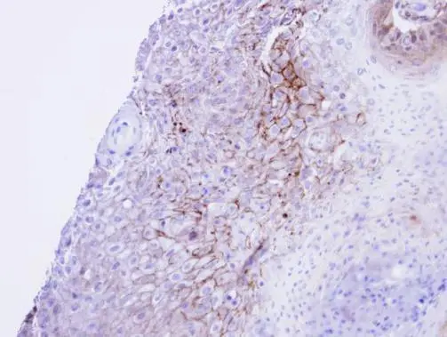
. CACNA1S was detected in paraffin-embedded section of human skeletal muscle tissue. Heat mediated antigen retrieval was performed in EDTA buffer (pH8.0, epitope retrieval solution). The tissue section was blocked with 10% goat serum. The tissue section was then incubated with 2microg/ml rabbit anti-CACNA1S Antibody (A03006-1) overnight at 4°C. Biotinylated goat anti-rabbit IgG was used as secondary antibody and incubated for 30 minutes at 37°C. The tissue section was developed using Strepavidin-Biotin-Complex (SABC) (Catalog # SA1022) with DAB as the chromogen.")
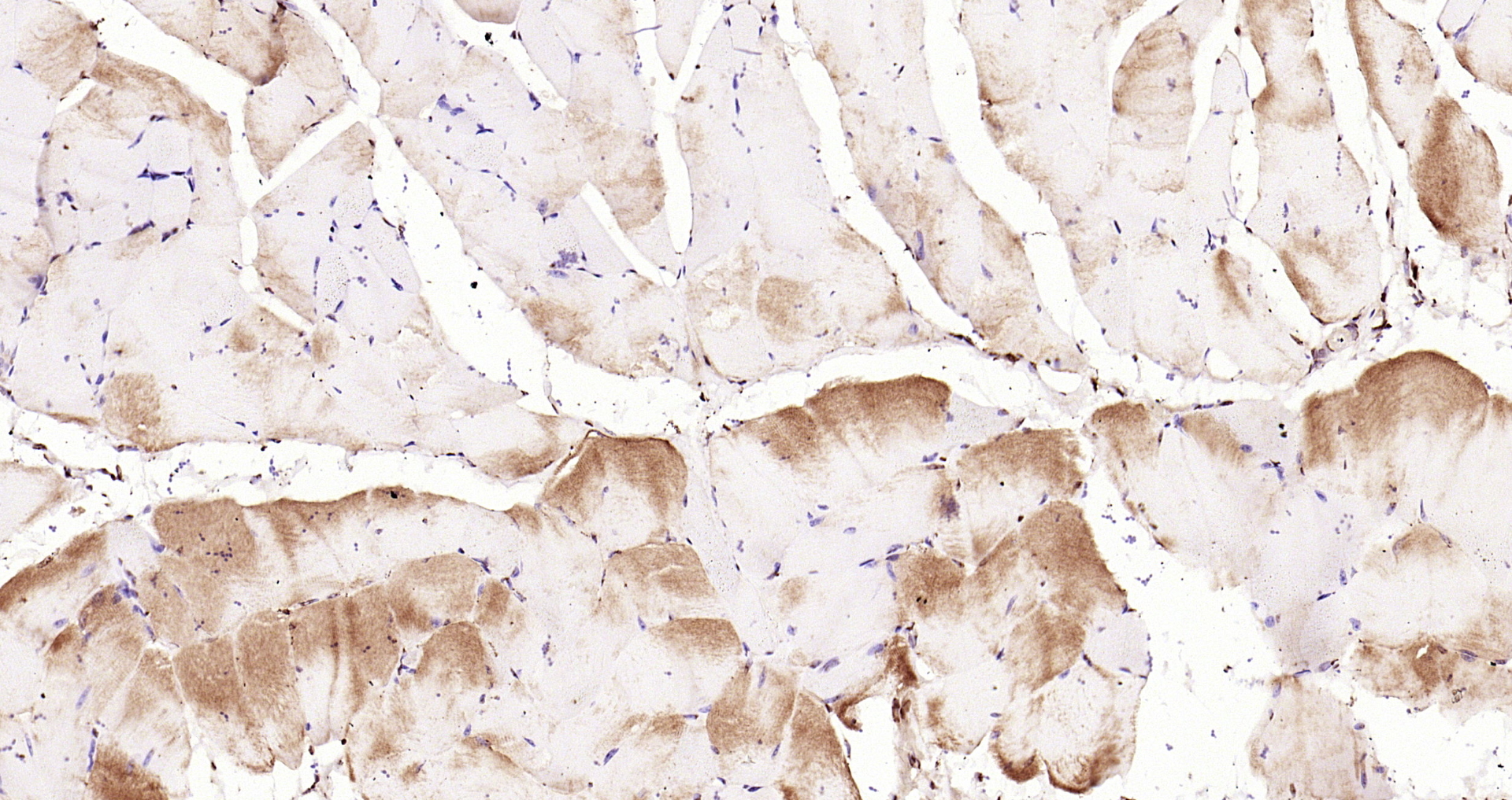
. CACNA1S was detected in paraffin-embedded section of rat skeletal muscle tissue. Heat mediated antigen retrieval was performed in EDTA buffer (pH8.0, epitope retrieval solution). The tissue section was blocked with 10% goat serum. The tissue section was then incubated with 2microg/ml rabbit anti-CACNA1S Antibody (A03006-1) overnight at 4°C. Biotinylated goat anti-rabbit IgG was used as secondary antibody and incubated for 30 minutes at 37°C. The tissue section was developed using Strepavidin-Biotin-Complex (SABC) (Catalog # SA1022) with DAB as the chromogen.")
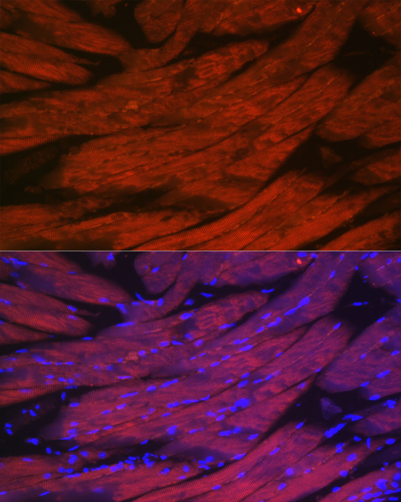
. Overlay histogram showing U937 cells stained with A03006-1 (Blue line). The cells were fixed with 4% paraformaldehyde and blocked with 10% normal goat serum. And then incubated with rabbit anti-CACNA1S Antibody (A03006-1, 1microg/1x106 cells) for 30 min at 20°C. DyLight®488 conjugated goat anti-rabbit IgG (BA1127, 5-10microg/1x106 cells) was used as secondary antibody for 30 minutes at 20°C. Isotype control antibody (Green line) was rabbit IgG (1microg/1x106) used under the same conditions. Unlabelled sample without incubation with primary antibody and secondary antibody (Red line) was used as a blank control.")
Figure 1. Western blot analysis of CACNA1S using anti-CACNA1S antibody (A03006-1). Electrophoresis was performed on a 5-20% SDS-PAGE gel at 70V (Stacking gel) / 90V (Resolving gel) for 2-3 hours. The sample well of each lane was loaded with 50ug of sample under reducing conditions. Lane 1: monkey skeletal muscle tissue lysates, Lane 2: rat skeletal muscle tissue lysates, Lane 3: mouse skeletal muscle tissue lysates. After Electrophoresis, proteins were transferred to a Nitrocellulose membrane at 150mA for 50-90 minutes. Blocked the membrane with 5% Non-fat Milk/ TBS for 1.5 hour at RT. The membrane was incubated with rabbit anti-CACNA1S antigen affinity purified polyclonal antibody (Catalog # A03006-1) at 0.5 microg/mL overnight at 4°C, then washed with TBS-0.1%Tween 3 times with 5 minutes each and probed with a goat anti-rabbit IgG-HRP secondary antibody at a dilution of 1:5000 for 1.5 hour at RT. The signal is developed using an Enhanced Chemiluminescent detection (ECL) kit (Catalog # EK1002) with Tanon 5200 system. A specific band was detected for CACNA1S at approximately 220KD. The expected band size for CACNA1S is at 220KD.
Anti-CACNA1S Antibody Picoband(r)
A03006-1-CARRIER-FREE
ApplicationsFlow Cytometry, Western Blot, ELISA, ImmunoHistoChemistry
Product group Antibodies
ReactivityHuman, Monkey, Mouse, Rat
TargetCACNA1S
Overview
- SupplierBoster Bio
- Product NameAnti-CACNA1S Antibody Picoband(r)
- Delivery Days Customer9
- ApplicationsFlow Cytometry, Western Blot, ELISA, ImmunoHistoChemistry
- CertificationResearch Use Only
- ClonalityPolyclonal
- Concentration500 ug/ml
- Gene ID779
- Target nameCACNA1S
- Target descriptioncalcium voltage-gated channel subunit alpha1 S
- Target synonymsCACNL1A3, CCHL1A3, CMYO18, CMYP18, Cav1.1, DHPRM, HOKPP, HOKPP1, MHS5, TTPP1, hypoPP, voltage-dependent L-type calcium channel subunit alpha-1S, calcium channel, voltage-dependent, L type, alpha 1S subunit, dihydropyridine receptor alpha 1 subunit, dihydropyridine-sensitive L-type calcium channel alpha-1 subunit, voltage-gated calcium channel subunit alpha Cav1.1
- HostRabbit
- IsotypeIgG
- Protein IDQ13698
- Protein NameVoltage-dependent L-type calcium channel subunit alpha-1S
- Scientific DescriptionBoster Bio Anti-CACNA1S Antibody Picoband® catalog # A03006-1. Tested in ELISA, Flow Cytometry, IHC, WB applications. This antibody reacts with Human, Monkey, Mouse, Rat. The brand Picoband indicates this is a premium antibody that guarantees superior quality, high affinity, and strong signals with minimal background in Western blot applications. Only our best-performing antibodies are designated as Picoband, ensuring unmatched performance.
- ReactivityHuman, Monkey, Mouse, Rat
- Storage Instruction-20°C,2°C to 8°C
- UNSPSC12352203
Related products
Product group Antibodies
Anti-CACNA1S Antibody107-10356
ApplicationsWestern Blot, ImmunoHistoChemistry, ImmunoHistoChemistry Paraffin
ReactivityHuman
TargetCACNA1S
- SizePrice
Product group Antibodies
References
Cav1.1 antibody [N2N3]GTX102941
ApplicationsWestern Blot, ImmunoHistoChemistry, ImmunoHistoChemistry Paraffin
ReactivityHuman
TargetCACNA1S
- SizePrice
Product group Antibodies
Anti-CACNA1S AntibodyA309295
ApplicationsImmunoFluorescence, ImmunoCytoChemistry
ReactivityMouse, Rat
- SizePrice
Product group Antibodies
References
CACNA1S Polyclonal AntibodyBS-9925R
ApplicationsImmunoFluorescence, ELISA, ImmunoCytoChemistry, ImmunoHistoChemistry, ImmunoHistoChemistry Paraffin
ReactivityBovine, Human, Mouse, Porcine, Rabbit, Rat
TargetCACNA1S
- SizePrice
Product group Antibodies

CACNA1S / Cav1.1 Antibody (aa1505-1646)LS-C373333
ApplicationsWestern Blot
ReactivityMouse
TargetCACNA1S
- SizePrice

Product group Antibodies
Anti-CACNA1S AntibodyHPA048892
ApplicationsImmunoHistoChemistry
ReactivityHuman
TargetCACNA1S
- SizePrice