


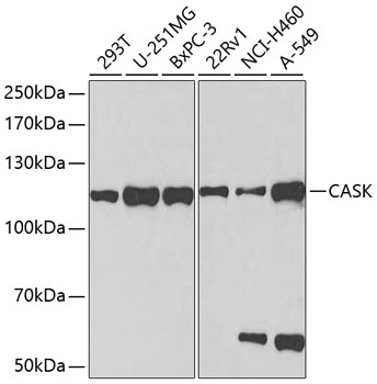
Figure 1. Western blot analysis of CASK using anti-CASK antibody (A02468-2). Electrophoresis was performed on a 5-20% SDS-PAGE gel at 70V (Stacking gel) / 90V (Resolving gel) for 2-3 hours. The sample well of each lane was loaded with 30 ug of sample under reducing conditions. Lane 1: human CACO-2 whole cell lysates, Lane 2: human MCF-7 whole cell lysates, Lane 3: rat kidney tissue lysates, Lane 4: rat brain tissue lysates, Lane 5: mouse kidney tissue lysates, Lane 6: mouse brain tissue lysates. After electrophoresis, proteins were transferred to a nitrocellulose membrane at 150 mA for 50-90 minutes. Blocked the membrane with 5% non-fat milk/TBS for 1.5 hour at RT. The membrane was incubated with rabbit anti-CASK antigen affinity purified polyclonal antibody (Catalog # A02468-2) at 0.5 microg/mL overnight at 4°C, then washed with TBS-0.1%Tween 3 times with 5 minutes each and probed with a goat anti-rabbit IgG-HRP secondary antibody at a dilution of 1:5000 for 1.5 hour at RT. The signal is developed using an Enhanced Chemiluminescent detection (ECL) kit (Catalog # EK1002) with Tanon 5200 system. A specific band was detected for CASK at approximately 120 kDa. The expected band size for CASK is at 120 kDa.
. Overlay histogram showing CACO-2 cells stained with A02468-2 (Blue line). To facilitate intracellular staining, cells were fixed with 4% paraformaldehyde and permeabilized with permeabilization buffer. The cells were blocked with 10% normal goat serum. And then incubated with rabbit anti-CASK Antibody (A02468-2, 1 microg/1x106 cells) for 30 min at 20°C. DyLight®488 conjugated goat anti-rabbit IgG (BA1127, 5-10 microg/1x106 cells) was used as secondary antibody for 30 minutes at 20°C. Isotype control antibody (Green line) was rabbit IgG (1 microg/1x106) used under the same conditions. Unlabelled sample without incubation with primary antibody and secondary antibody (Red line) was used as a blank control.")
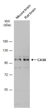
Figure 1. Western blot analysis of CASK using anti-CASK antibody (A02468-2). Electrophoresis was performed on a 5-20% SDS-PAGE gel at 70V (Stacking gel) / 90V (Resolving gel) for 2-3 hours. The sample well of each lane was loaded with 30 ug of sample under reducing conditions. Lane 1: human CACO-2 whole cell lysates, Lane 2: human MCF-7 whole cell lysates, Lane 3: rat kidney tissue lysates, Lane 4: rat brain tissue lysates, Lane 5: mouse kidney tissue lysates, Lane 6: mouse brain tissue lysates. After electrophoresis, proteins were transferred to a nitrocellulose membrane at 150 mA for 50-90 minutes. Blocked the membrane with 5% non-fat milk/TBS for 1.5 hour at RT. The membrane was incubated with rabbit anti-CASK antigen affinity purified polyclonal antibody (Catalog # A02468-2) at 0.5 microg/mL overnight at 4°C, then washed with TBS-0.1%Tween 3 times with 5 minutes each and probed with a goat anti-rabbit IgG-HRP secondary antibody at a dilution of 1:5000 for 1.5 hour at RT. The signal is developed using an Enhanced Chemiluminescent detection (ECL) kit (Catalog # EK1002) with Tanon 5200 system. A specific band was detected for CASK at approximately 120 kDa. The expected band size for CASK is at 120 kDa.
Anti-CASK Antibody Picoband(r)
A02468-2-CARRIER-FREE
ApplicationsFlow Cytometry, Western Blot, ELISA
Product group Antibodies
ReactivityHuman, Mouse, Rat
TargetCASK
Overview
- SupplierBoster Bio
- Product NameAnti-CASK Antibody Picoband(r)
- Delivery Days Customer9
- ApplicationsFlow Cytometry, Western Blot, ELISA
- CertificationResearch Use Only
- ClonalityPolyclonal
- Concentration500 ug/ml
- Gene ID8573
- Target nameCASK
- Target descriptioncalcium/calmodulin dependent serine protein kinase
- Target synonymsCAGH39, CAMGUK, CMG, FGS4, LIN2, MICPCH, MRXSNA, TNRC8, hCASK, peripheral plasma membrane protein CASK, calcium/calmodulin-dependent serin protein kinase, calcium/calmodulin-dependent serine protein kinase (MAGUK family), calcium/calmodulin-dependent serine protein kinase membrane-associated guanylate kinase, protein lin-2 homolog, trinucleotide repeat containing 8
- HostRabbit
- IsotypeIgG
- Protein IDO14936
- Protein NamePeripheral plasma membrane protein CASK
- Scientific DescriptionBoster Bio Anti-CASK Antibody Picoband® catalog # A02468-2. Tested in ELISA, Flow Cytometry, WB applications. This antibody reacts with Human, Mouse, Rat. The brand Picoband indicates this is a premium antibody that guarantees superior quality, high affinity, and strong signals with minimal background in Western blot applications. Only our best-performing antibodies are designated as Picoband, ensuring unmatched performance.
- ReactivityHuman, Mouse, Rat
- Storage Instruction-20°C,2°C to 8°C
- UNSPSC12352203
Related products
Product group Antibodies
Anti-CASK Antibody144-02501
ApplicationsImmunoFluorescence, Western Blot
ReactivityHuman, Mouse
TargetCASK
- SizePrice
Product group Antibodies
Anti-CASK AntibodyA14013
ApplicationsImmunoFluorescence, Western Blot, ImmunoCytoChemistry
ReactivityHuman
- SizePrice
Product group Antibodies

CASK AntibodyLS-C831944
ApplicationsELISA, ImmunoHistoChemistry
ReactivityHuman, Mouse, Rat
TargetCASK
- SizePrice
Product group Antibodies
CASK Polyclonal AntibodyBS-11338R
ApplicationsImmunoFluorescence, ELISA, ImmunoCytoChemistry, ImmunoHistoChemistry, ImmunoHistoChemistry Frozen, ImmunoHistoChemistry Paraffin
ReactivityBovine, Chicken, Equine, Human, Mouse, Porcine, Rabbit, Rat, Sheep
- SizePrice
Product group Antibodies
Cask Polyclonal AntibodyCAC11784
ApplicationsELISA, ImmunoHistoChemistry
TargetCASK
- SizePrice
Product group Antibodies
CASK AntibodyCSB-PA004539LA01HU
ApplicationsELISA, ImmunoHistoChemistry
ReactivityHuman
TargetCASK
- SizePrice

Product group Antibodies
Anti-CASK AntibodyHPA023857
ApplicationsImmunoCytoChemistry, ImmunoHistoChemistry
ReactivityHuman
TargetCASK
- SizePrice
Product group Antibodies
CASK antibodyGTX111513
ApplicationsImmunoFluorescence, Western Blot, ImmunoCytoChemistry, ImmunoHistoChemistry, ImmunoHistoChemistry Paraffin
ReactivityHuman, Mouse, Rat
TargetCASK
- SizePrice
Product group Antibodies
Anti-CASK AntibodyCAB2501
ApplicationsImmunoFluorescence, Western Blot, ELISA, ImmunoCytoChemistry
ReactivityHuman
TargetCASK
- SizePrice