

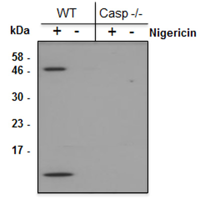
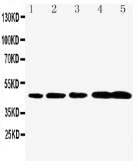
Mouse Caspase-1 (p10) is detected by immunoblotting using anti-Caspase-1 (p10) (mouse), mAb (Casper-2) (Prod. No AG-20B-0044). Method: Caspase-1 was analyzed by Western blot in supernatants of differentiated bone marrow-derived dendritic cells (BMD
Mouse Caspase-1 (p10) is detected by immunoblotting using anti-Caspase-1 (p10) (mouse), mAb (Casper-2) (Prod. No AG-20B-0044). Method: Caspase-1 was analyzed by Western blot in supernatants of differentiated bone marrow-derived dendritic cells (BMD
anti-Caspase-1 (p10) (mouse), mAb (Casper-2)
AG-20B-0044
ApplicationsWestern Blot
Product group Antibodies
ReactivityMouse
TargetCasp1
Overview
- SupplierAdipoGen Life Sciences
- Product Nameanti-Caspase-1 (p10) (mouse), mAb (Casper-2)
- Delivery Days Customer10
- ApplicationsWestern Blot
- CertificationResearch Use Only
- ClonalityMonoclonal
- Clone IDCasper-2
- Concentration1 mg/ml
- Estimated Purity>95%
- Gene ID12362
- Target nameCasp1
- Target descriptioncaspase 1
- Target synonymsICE, Il1bc, caspase-1, CASP-1, IL-1 beta-converting enzyme, IL-1B converting enzyme, IL-1BC, interleukin-1 beta convertase, interleukin-1 beta-converting enzyme, p45
- HostMouse
- IsotypeIgG2a
- Protein IDP29452
- Protein NameCaspase-1
- Scientific DescriptionCaspase-1 is the best-described inflammatory caspase. It processes the cytokines interleukin-1beta (IL-1beta) and IL-18 and induces pyroptotic cell death. Caspase-1 is activated by multiprotein complexes called inflammasomes in response to numerous stimuli that are detected through distinct inflammasomes. NLRC4 responds to cytosolic flagellin, murine NLRP1b responds to anthrax lethal toxin, AIM2 responds to cytosolic DNA and NLRP3 responds to a variety of agonists including crystals. - Monoclonal Antibody. Recognizes endogenous full-length and activated (p10 fragment) mouse caspase-1. Isotype: Mouse IgG2a. Clone: Casper-2. Applications: WB. Liquid. In PBS containing 10% glycerol and 0.02% sodium azide. Caspase-1 is the best-described inflammatory caspase. It processes the cytokines interleukin-1beta (IL-1beta) and IL-18 and induces pyroptotic cell death. Caspase-1 is activated by multiprotein complexes called inflammasomes in response to numerous stimuli that are detected through distinct inflammasomes. NLRC4 responds to cytosolic flagellin, murine NLRP1b responds to anthrax lethal toxin, AIM2 responds to cytosolic DNA and NLRP3 responds to a variety of agonists including crystals.
- ReactivityMouse
- Storage Instruction-20°C,2°C to 8°C
- UNSPSC41116161
MSDS
Related products
Product group Antibodies
anti-Caspase-1 (p20) (mouse), mAb (Casper-1)AG-20B-0042
ApplicationsImmunoPrecipitation, Western Blot, ImmunoHistoChemistry
ReactivityMouse
TargetCasp1
- SizePrice
Product group Antibodies
ApplicationsImmunoPrecipitation, Western Blot, ImmunoHistoChemistry
ReactivityMouse
TargetCasp1
- SizePrice
Product group Antibodies
ApplicationsWestern Blot
ReactivityMouse
TargetCasp1
- SizePrice
Product group Antibodies
anti-Caspase-1 (p20) (human), mAb (Bally-1)AG-20B-0048
ApplicationsWestern Blot
ReactivityHuman
TargetCASP1
- SizePrice
Product group Antibodies
ApplicationsWestern Blot
ReactivityHuman
TargetCASP1
- SizePrice
Product group Antibodies
Caspase-1 p20 Polyclonal AntibodyBS-10442R
ApplicationsFlow Cytometry, ImmunoFluorescence, Western Blot, ELISA, ImmunoCytoChemistry, ImmunoHistoChemistry, ImmunoHistoChemistry Frozen, ImmunoHistoChemistry Paraffin
ReactivityHuman, Mouse, Rat
TargetCasp1
- SizePrice
Product group Antibodies
ApplicationsImmunoPrecipitation, Western Blot, ImmunoCytoChemistry, ImmunoHistoChemistry
ReactivityMouse, Rat
TargetCasp1
- SizePrice
Product group Antibodies
References
Caspase 1 p20 subunit antibodyGTX11701
ApplicationsWestern Blot
ReactivityMouse, Rat
TargetCasp1
- SizePrice

Product group Antibodies
Anti-Caspase-1(P20)/CASP1 Antibody Picoband(r)PA1440-1-CARRIER-FREE
ApplicationsWestern Blot
ReactivityMouse, Rat
TargetCasp1
- SizePrice