


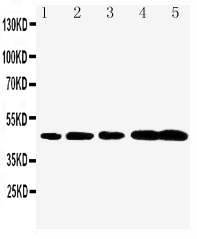
Figure 1. Western blot analysis of CASP1 using anti-CASP1 antibody (PA1440-1). Electrophoresis was performed on a 5-20% SDS-PAGE gel at 70V (Stacking gel) / 90V (Resolving gel) for 2-3 hours. The sample well of each lane was loaded with 30 ug of sample under reducing conditions. Lane 1: rat thymus tissue lysates, Lane 2: rat C6 whole cell lysates, Lane 3: mouse thymus tissue lysates. After electrophoresis, proteins were transferred to a nitrocellulose membrane at 150 mA for 50-90 minutes. Blocked the membrane with 5% non-fat milk/TBS for 1.5 hour at RT. The membrane was incubated with rabbit anti-CASP1 antigen affinity purified polyclonal antibody (Catalog # PA1440-1) at 0.5 microg/mL overnight at 4°C, then washed with TBS-0.1%Tween 3 times with 5 minutes each and probed with a goat anti-rabbit IgG-HRP secondary antibody at a dilution of 1:5000 for 1.5 hour at RT. The signal is developed using an Enhanced Chemiluminescent detection (ECL) kit (Catalog # EK1002) with Tanon 5200 system. A specific band was detected for CASP1 at approximately 46 kDa. The expected band size for CASP1 is at 46 kDa.
Figure 1. Western blot analysis of CASP1 using anti-CASP1 antibody (PA1440-1). Electrophoresis was performed on a 5-20% SDS-PAGE gel at 70V (Stacking gel) / 90V (Resolving gel) for 2-3 hours. The sample well of each lane was loaded with 30 ug of sample under reducing conditions. Lane 1: rat thymus tissue lysates, Lane 2: rat C6 whole cell lysates, Lane 3: mouse thymus tissue lysates. After electrophoresis, proteins were transferred to a nitrocellulose membrane at 150 mA for 50-90 minutes. Blocked the membrane with 5% non-fat milk/TBS for 1.5 hour at RT. The membrane was incubated with rabbit anti-CASP1 antigen affinity purified polyclonal antibody (Catalog # PA1440-1) at 0.5 microg/mL overnight at 4°C, then washed with TBS-0.1%Tween 3 times with 5 minutes each and probed with a goat anti-rabbit IgG-HRP secondary antibody at a dilution of 1:5000 for 1.5 hour at RT. The signal is developed using an Enhanced Chemiluminescent detection (ECL) kit (Catalog # EK1002) with Tanon 5200 system. A specific band was detected for CASP1 at approximately 46 kDa. The expected band size for CASP1 is at 46 kDa.
Anti-Caspase-1(P20)/CASP1 Antibody Picoband(r)
PA1440-1
ApplicationsWestern Blot
Product group Antibodies
ReactivityMouse, Rat
TargetCasp1
Overview
- SupplierBoster Bio
- Product NameAnti-Caspase-1(P20)/CASP1 Antibody Picoband(r)
- Delivery Days Customer9
- Application Supplier NoteTested Species: In-house tested species with positive results. Predicted Species: Species predicted to be fit for the product based on sequence similarities. By Heat: Boiling the paraffin sections in 10mM citrate buffer, pH6.0, for 20mins is required for the staining of formalin/paraffin sections. Other applications have not been tested. Optimal dilutions should be determined by end users.
- ApplicationsWestern Blot
- Applications SupplierWB
- CertificationResearch Use Only
- ClonalityPolyclonal
- Concentration500 ug/ml
- Gene ID12362
- Target nameCasp1
- Target descriptioncaspase 1
- Target synonymsICE, Il1bc, caspase-1, CASP-1, IL-1 beta-converting enzyme, IL-1B converting enzyme, IL-1BC, interleukin-1 beta convertase, interleukin-1 beta-converting enzyme, p45
- HostRabbit
- IsotypeIgG
- Protein IDP29452
- Protein NameCaspase-1
- Scientific DescriptionBoster Bio Anti-Caspase-1 (P20)/CASP1 Antibody catalog # PA1440-1. Tested in WB applications. This antibody reacts with Mouse, Rat. The brand Picoband indicates this is a premium antibody that guarantees superior quality, high affinity, and strong signals with minimal background in Western blot applications. Only our best-performing antibodies are designated as Picoband, ensuring unmatched performance.
- ReactivityMouse, Rat
- Storage Instruction-20°C,2°C to 8°C
- UNSPSC12352203
References
- Yang L, Xing W, Shi Y, et al. Stress-induced NLRP3 inflammasome activation and myelin alterations in the hippocampus of PTSD rats. Neuroscience. 2024,555:156-166. doi: 10.1016/j.neuroscience.2024.07.028Read this paper
- Jia R, Ma H, Hao H, et al. Metformin inhibits activation of NLRP3 inflammasome and inflammatory response in preeclamptic rats. Gene. 2024,919:148509. doi: 10.1016/j.gene.2024.148509Read this paper
- Fan X, Zang C, Lao K, et al. Neuroprotective effects of tetramethylpyrazine on spinal cord injury-Related neuroinflammation mediated by P2X7R/NLRP3 interaction. Eur J Pharmacol. 2024,964:176267. doi: 10.1016/j.ejphar.2023.176267Read this paper
- Liu X, Li H, Feng Y, et al. Resatorvid alleviates experimental inflammatory TMJOA by restraining chondrocyte pyroptosis and synovial inflammation. Arthritis Res Ther. 2023,25(1):230. doi: 10.1186/s13075-023-03214-4Read this paper
- Liu X, Li Y, Zhao J, et al. Pyroptosis of chondrocytes activated by synovial inflammation accelerates TMJ osteoarthritis cartilage degeneration via ROS/NLRP3 signaling. Int Immunopharmacol. 2023,124(Pt A):110781. doi: 10.1016/j.intimp.2023.110781Read this paper
- Yang R, Wang Y, Mehmood S, et al. Polysaccharides from Armillariella tabescens mycelia mitigate DSS-induced ulcerative colitis via modulating intestinal microbiota in mice. Int J Biol Macromol. 2023,245:125538. doi: 10.1016/j.ijbiomac.2023.125538Read this paper
- Yang R, Chen J, Jia Q, et al. Epigallocatechin-3-gallate ameliorates renal endoplasmic reticulum stress-mediated inflammation in type 2 diabetic rats. Exp Biol Med (Maywood). 2022,247(16):1410-1419. doi: 10.1177/15353702221106479Read this paper
- Zhang X, Gao R, Zhou Z, et al. Uncovering the mechanism of Huanglian-Wuzhuyu herb pair in treating nonalcoholic steatohepatitis based on network pharmacology and experimental validation. J Ethnopharmacol. 2022,296:115405. doi: 10.1016/j.jep.2022.115405Read this paper
- Jiang Y, Liu H, Yu H, et al. Circular RNA Calm4 Regulates Hypoxia-Induced Pulmonary Arterial Smooth Muscle Cells Pyroptosis via the Circ-Calm4/miR-124-3p/PDCD6 Axis. Arterioscler Thromb Vasc Biol. 2021,41(5):1675-1693. doi: 10.1161/ATVBAHA.120.315525Read this paper
- Zhang XL, Huang WM, Tang PC, et al. Anti-inflammatory and neuroprotective effects of natural cordycepin in rotenone-induced PD models through inhibiting Drp1-mediated mitochondrial fission. Neurotoxicology. 2021,84:1-13. doi: 10.1016/j.neuro.2021.02.002Read this paper
Datasheet
MSDS
Related products
Product group Antibodies
ApplicationsImmunoPrecipitation, Western Blot, ImmunoCytoChemistry, ImmunoHistoChemistry
ReactivityMouse, Rat
TargetCasp1
- SizePrice
Product group Antibodies
anti-Caspase-1 (p20) (mouse), mAb (Casper-1)AG-20B-0042
ApplicationsImmunoPrecipitation, Western Blot, ImmunoHistoChemistry
ReactivityMouse
TargetCasp1
- SizePrice
Product group Antibodies
Anti-Caspase-1(P20)/CASP1 Antibody Picoband(r)PA1440-1-CARRIER-FREE
ApplicationsWestern Blot
ReactivityMouse, Rat
TargetCasp1
- SizePrice
Product group Antibodies
References
Caspase 1 p20 subunit antibodyGTX11701
ApplicationsWestern Blot
ReactivityMouse, Rat
TargetCasp1
- SizePrice
Product group Antibodies
Caspase-1 p20 Polyclonal AntibodyBS-10442R
ApplicationsFlow Cytometry, ImmunoFluorescence, Western Blot, ELISA, ImmunoCytoChemistry, ImmunoHistoChemistry, ImmunoHistoChemistry Frozen, ImmunoHistoChemistry Paraffin
ReactivityHuman, Mouse, Rat
TargetCasp1
- SizePrice