


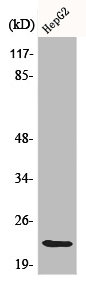
Figure 1. Western blot analysis of Caveolin-1/CAV1 using anti-Caveolin-1/CAV1 antibody (PB9165). Electrophoresis was performed on a 5-20% SDS-PAGE gel at 70V (Stacking gel) / 90V (Resolving gel) for 2-3 hours. The sample well of each lane was loaded with 30 ug of sample under reducing conditions. Lane 1: human Hela whole cell lysates, Lane 2: human A549 whole cell lysates, Lane 3: human placenta tissue lysates, Lane 4: human A431 whole cell lysates, Lane 5: human HL-60 whole cell lysates, Lane 6: rat ovary tissue lysates, Lane 7: rat heart tissue lysates, Lane 8: mouse ovary tissue lysates, Lane 9: mouse heart tissue lysates. After electrophoresis, proteins were transferred to a nitrocellulose membrane at 150 mA for 50-90 minutes. Blocked the membrane with 5% non-fat milk/TBS for 1.5 hour at RT. The membrane was incubated with rabbit anti-Caveolin-1/CAV1 antigen affinity purified polyclonal antibody (Catalog # PB9165) at 0.5 microg/mL overnight at 4°C, then washed with TBS-0.1%Tween 3 times with 5 minutes each and probed with a goat anti-rabbit IgG-HRP secondary antibody at a dilution of 1:5000 for 1.5 hour at RT. The signal is developed using an Enhanced Chemiluminescent detection (ECL) kit (Catalog # EK1002) with Tanon 5200 system. A specific band was detected for Caveolin-1/CAV1 at approximately 22 kDa. The expected band size for Caveolin-1/CAV1 is at 20 kDa.
. Caveolin-1/CAV1 was detected in a paraffin-embedded section of human glioma tissue. Heat mediated antigen retrieval was performed in EDTA buffer (pH 8.0, epitope retrieval solution). The tissue section was blocked with 10% goat serum. The tissue section was then incubated with 2 microg/ml rabbit anti-Caveolin-1/CAV1 Antibody (PB9165) overnight at 4°C. Peroxidase Conjugated Goat Anti-rabbit IgG was used as secondary antibody and incubated for 30 minutes at 37°C. The tissue section was developed using HRP Conjugated Rabbit IgG Super Vision Assay Kit (Catalog # SV0002) with DAB as the chromogen.")
. Caveolin-1/CAV1 was detected in a paraffin-embedded section of human meningeoma tissue. Heat mediated antigen retrieval was performed in EDTA buffer (pH 8.0, epitope retrieval solution). The tissue section was blocked with 10% goat serum. The tissue section was then incubated with 2 microg/ml rabbit anti-Caveolin-1/CAV1 Antibody (PB9165) overnight at 4°C. Peroxidase Conjugated Goat Anti-rabbit IgG was used as secondary antibody and incubated for 30 minutes at 37°C. The tissue section was developed using HRP Conjugated Rabbit IgG Super Vision Assay Kit (Catalog # SV0002) with DAB as the chromogen.")
. Caveolin-1/CAV1 was detected in a paraffin-embedded section of mouse brain tissue. Heat mediated antigen retrieval was performed in EDTA buffer (pH 8.0, epitope retrieval solution). The tissue section was blocked with 10% goat serum. The tissue section was then incubated with 2 microg/ml rabbit anti-Caveolin-1/CAV1 Antibody (PB9165) overnight at 4°C. Peroxidase Conjugated Goat Anti-rabbit IgG was used as secondary antibody and incubated for 30 minutes at 37°C. The tissue section was developed using HRP Conjugated Rabbit IgG Super Vision Assay Kit (Catalog # SV0002) with DAB as the chromogen.")
. Caveolin-1/CAV1 was detected in a frozen section of human placenta tissue. The tissue section was blocked with 10% goat serum. The tissue section was then incubated with 2 microg/ml rabbit anti-Caveolin-1/CAV1 Antibody (PB9165) overnight at 4°C. Peroxidase Conjugated Goat Anti-rabbit IgG was used as secondary antibody and incubated for 30 minutes at 37°C. The tissue section was developed using HRP Conjugated Rabbit IgG Super Vision Assay Kit (Catalog # SV0002) with DAB as the chromogen.")
. Caveolin-1/CAV1 was detected in a paraffin-embedded section of human glioma tissue. Heat mediated antigen retrieval was performed in EDTA buffer (pH 8.0, epitope retrieval solution). The tissue section was blocked with 10% goat serum. The tissue section was then incubated with 5 microg/mL rabbit anti-Caveolin-1/CAV1 Antibody (PB9165) overnight at 4°C. Cy3 Conjugated Goat Anti-Rabbit IgG (BA1032) was used as secondary antibody at 1:500 dilution and incubated for 30 minutes at 37°C. The section was counterstained with DAPI. Visualize using a fluorescence microscope and filter sets appropriate for the label used.")
. Overlay histogram showing U87 cells stained with PB9165 (Blue line). To facilitate intracellular staining, cells were fixed with 4% paraformaldehyde and permeabilized with permeabilization buffer. The cells were blocked with 10% normal goat serum. And then incubated with rabbit anti-Caveolin-1/CAV1 Antibody (PB9165, 1 microg/1x106 cells) for 30 min at 20°C. DyLight®488 conjugated goat anti-rabbit IgG (BA1127, 5-10 microg/1x106 cells) was used as secondary antibody for 30 minutes at 20°C. Isotype control antibody (Green line) was rabbit IgG (1 microg/1x106) used under the same conditions. Unlabelled sample (Red line) was also used as a control.")
Figure 1. Western blot analysis of Caveolin-1/CAV1 using anti-Caveolin-1/CAV1 antibody (PB9165). Electrophoresis was performed on a 5-20% SDS-PAGE gel at 70V (Stacking gel) / 90V (Resolving gel) for 2-3 hours. The sample well of each lane was loaded with 30 ug of sample under reducing conditions. Lane 1: human Hela whole cell lysates, Lane 2: human A549 whole cell lysates, Lane 3: human placenta tissue lysates, Lane 4: human A431 whole cell lysates, Lane 5: human HL-60 whole cell lysates, Lane 6: rat ovary tissue lysates, Lane 7: rat heart tissue lysates, Lane 8: mouse ovary tissue lysates, Lane 9: mouse heart tissue lysates. After electrophoresis, proteins were transferred to a nitrocellulose membrane at 150 mA for 50-90 minutes. Blocked the membrane with 5% non-fat milk/TBS for 1.5 hour at RT. The membrane was incubated with rabbit anti-Caveolin-1/CAV1 antigen affinity purified polyclonal antibody (Catalog # PB9165) at 0.5 microg/mL overnight at 4°C, then washed with TBS-0.1%Tween 3 times with 5 minutes each and probed with a goat anti-rabbit IgG-HRP secondary antibody at a dilution of 1:5000 for 1.5 hour at RT. The signal is developed using an Enhanced Chemiluminescent detection (ECL) kit (Catalog # EK1002) with Tanon 5200 system. A specific band was detected for Caveolin-1/CAV1 at approximately 22 kDa. The expected band size for Caveolin-1/CAV1 is at 20 kDa.
Anti-Caveolin-1/CAV1 Antibody Picoband(r)
PB9165-CARRIER-FREE
ApplicationsFlow Cytometry, ImmunoFluorescence, Western Blot, ImmunoHistoChemistry, ImmunoHistoChemistry Frozen
Product group Antibodies
ReactivityHuman, Mouse, Rat
TargetCAV1
Overview
- SupplierBoster Bio
- Product NameAnti-Caveolin-1/CAV1 Antibody Picoband(r)
- Delivery Days Customer9
- Application Supplier NoteWB: The detection limit for Caveolin-1 is approximately 0.25ng/lane under reducing conditions. Tested Species: In-house tested species with positive results. By Heat: Boiling the paraffin sections in 10mM citrate buffer, pH6.0, for 20mins is required for the staining of formalin/paraffin sections. Other applications have not been tested. Optimal dilutions should be determined by end users.
- ApplicationsFlow Cytometry, ImmunoFluorescence, Western Blot, ImmunoHistoChemistry, ImmunoHistoChemistry Frozen
- CertificationResearch Use Only
- ClonalityPolyclonal
- Concentration500 ug/ml
- Gene ID857
- Target nameCAV1
- Target descriptioncaveolin 1
- Target synonymsBSCL3, CGL3, LCCNS, MSTP085, PPH3, VIP21, caveolin-1, caveolin 1, caveolae protein, 22kDa, cell growth-inhibiting protein 32
- HostRabbit
- IsotypeIgG
- Protein IDQ03135
- Protein NameCaveolin-1
- Scientific DescriptionBoster Bio Anti-Caveolin-1/CAV1 Antibody Picoband® catalog # PB9165. Tested in Flow Cytometry, IF, IHC, IHC-F, WB applications. This antibody reacts with Human, Mouse, Rat. The brand Picoband indicates this is a premium antibody that guarantees superior quality, high affinity, and strong signals with minimal background in Western blot applications. Only our best-performing antibodies are designated as Picoband, ensuring unmatched performance.
- ReactivityHuman, Mouse, Rat
- Storage Instruction-20°C,2°C to 8°C
- UNSPSC12352203
Related products
Product group Antibodies
CAV1 AntibodyCSB-PA001332
ApplicationsWestern Blot, ELISA
ReactivityHuman, Mouse, Rat
TargetCAV1
- SizePrice
Product group Antibodies
Anti-Caveolin-1 AntibodyA82916
ApplicationsFlow Cytometry, ImmunoFluorescence, Western Blot, ELISA, ImmunoHistoChemistry
ReactivityHuman
- SizePrice
Product group Antibodies

Goat anti-Caveolin 1, BiotinylatedEB06817-B
ApplicationsWestern Blot, ELISA, ImmunoCytoChemistry
ReactivityHuman
TargetCAV1
- SizePrice

Product group Antibodies
Anti-CAV1-25ulHPA049326
ApplicationsWestern Blot, ImmunoHistoChemistry
ReactivityHuman
- SizePrice
Product group Antibodies

CAV1 / Caveolin 1 AntibodyLS-C402963
ApplicationsWestern Blot, ELISA, ImmunoHistoChemistry
ReactivityHuman, Mouse, Rat
TargetCAV1
- SizePrice
Product group Antibodies
ApplicationsWestern Blot, ImmunoHistoChemistry
ReactivityMouse, Porcine
TargetCAV1
- SizePrice

Product group Antibodies
References
Caveolin-1 Polyclonal AntibodyBS-1453R
ApplicationsFlow Cytometry, ImmunoFluorescence, Western Blot, ELISA, ImmunoCytoChemistry, ImmunoHistoChemistry, ImmunoHistoChemistry Frozen, ImmunoHistoChemistry Paraffin
ReactivityBovine, Canine, Equine, Human, Mouse, Porcine, Rabbit, Rat, Sheep
TargetCAV1
- SizePrice
![Wild-type (WT) and Caveolin 1 knockout (KO) HeLa cell extracts (30 μg) were separated by 12% SDS-PAGE, and the membrane was blotted with Caveolin 1 antibody [N1N3] (GTX100205) diluted at 1:500. The HRP-conjugated anti-rabbit IgG antibody (GTX213110-01) was used to detect the primary antibody.](https://www.genetex.com/upload/website/prouct_img/normal/GTX100205/GTX100205_39274_20170525_WB_KO_watermark_w_23053123_475.webp)
Product group Antibodies
Caveolin 1 antibody [N1N3]GTX100205
ApplicationsImmunoFluorescence, ImmunoPrecipitation, Western Blot, ImmunoCytoChemistry, ImmunoHistoChemistry, ImmunoHistoChemistry Paraffin
ReactivityHuman, Mouse, Rat
TargetCAV1
- SizePrice