

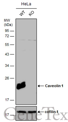
Wild-type (WT) and Caveolin 1 knockout (KO) HeLa cell extracts (30 μg) were separated by 12% SDS-PAGE, and the membrane was blotted with Caveolin 1 antibody [N1N3] (GTX100205) diluted at 1:500. The HRP-conjugated anti-rabbit IgG antibody (GTX213110-01) was used to detect the primary antibody.
![Caveolin 1 antibody [N1N3] detects Caveolin 1 protein at membrane by immunofluorescent analysis. Sample: A431 cells were fixed in ice-cold MeOH for 5 min. Green: Caveolin 1 protein stained by Caveolin 1 antibody [N1N3] (GTX100205) diluted at 1:500. Blue: Hoechst 33343 staining.](https://www.genetex.com/upload/website/prouct_img/normal/GTX100205/GTX100205_39274_IFA_w_23053123_803.webp "Caveolin 1 antibody [N1N3] detects Caveolin 1 protein at membrane by immunofluorescent analysis. Sample: A431 cells were fixed in ice-cold MeOH for 5 min. Green: Caveolin 1 protein stained by Caveolin 1 antibody [N1N3] (GTX100205) diluted at 1:500. Blue: Hoechst 33343 staining.")
 were separated by 12% SDS-PAGE, and the membrane was blotted with Caveolin 1 antibody (GTX100205) diluted at a dilution of 1:500. The HRP-conjugated anti-rabbit IgG antibody (GTX213110-01) was used to detect the primary antibody.")
![Immunohistochemical microscopy analysis of paraffin-embedded lung carcinoma cell line xenograft tissue using Caveolin-1 antibody [N1N3] (GTX100205) (1:100).
Antigen Retrieval: Trilogy? (EDTA based, pH 8.0) buffer, 15min](https://www.genetex.com/upload/website/prouct_img/normal/GTX100205/GTX100205_IHC_w_23053123_317.webp "Immunohistochemical microscopy analysis of paraffin-embedded lung carcinoma cell line xenograft tissue using Caveolin-1 antibody [N1N3] (GTX100205) (1:100).
Antigen Retrieval: Trilogy? (EDTA based, pH 8.0) buffer, 15min")
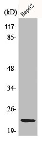
![Caveolin 1 antibody [N1N3] detects Caveolin 1 protein by western blot analysis. Mouse tissue extracts (50 μg) was separated by 12% SDS-PAGE, and the membrane was blotted with Caveolin 1 antibody [N1N3] (GTX100205) diluted at 1:500. The HRP-conjugated anti-rabbit IgG antibody (GTX213110-01) was used to detect the primary antibody.](https://www.genetex.com/upload/website/prouct_img/normal/GTX100205/GTX100205_39274_20151029_WB_M_lung_w_23053123_423.webp "Caveolin 1 antibody [N1N3] detects Caveolin 1 protein by western blot analysis. Mouse tissue extracts (50 μg) was separated by 12% SDS-PAGE, and the membrane was blotted with Caveolin 1 antibody [N1N3] (GTX100205) diluted at 1:500. The HRP-conjugated anti-rabbit IgG antibody (GTX213110-01) was used to detect the primary antibody.")
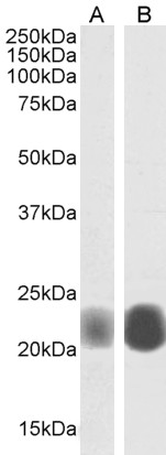
![Immunoprecipitation of Caveolin 1 protein from A549 membrane extracts using 5 μg of Caveolin 1 antibody [N1N3] (GTX100205). Western blot analysis was performed using Caveolin 1 antibody [N1N3] (GTX100205) diluted at 1:500. EasyBlot anti-Rabbit IgG (GTX221666-01) was used as a secondary reagent.](https://www.genetex.com/upload/website/prouct_img/normal/GTX100205/GTX100205_39274_20150106_IP_w_23060100_899.webp "Immunoprecipitation of Caveolin 1 protein from A549 membrane extracts using 5 μg of Caveolin 1 antibody [N1N3] (GTX100205). Western blot analysis was performed using Caveolin 1 antibody [N1N3] (GTX100205) diluted at 1:500. EasyBlot anti-Rabbit IgG (GTX221666-01) was used as a secondary reagent.")


Wild-type (WT) and Caveolin 1 knockout (KO) HeLa cell extracts (30 μg) were separated by 12% SDS-PAGE, and the membrane was blotted with Caveolin 1 antibody [N1N3] (GTX100205) diluted at 1:500. The HRP-conjugated anti-rabbit IgG antibody (GTX213110-01) was used to detect the primary antibody.
Caveolin 1 antibody [N1N3]
GTX100205
ApplicationsImmunoFluorescence, ImmunoPrecipitation, Western Blot, ImmunoCytoChemistry, ImmunoHistoChemistry, ImmunoHistoChemistry Paraffin
Product group Antibodies
ReactivityHuman, Mouse, Rat
TargetCAV1
Overview
- SupplierGeneTex
- Product NameCaveolin 1 antibody [N1N3]
- Delivery Days Customer9
- Application Supplier NoteWB: 1:500-1:3000. ICC/IF: 1:100-1:1000. IHC-P: 1:100-1:1000. IP: 1:100-1:500. *Optimal dilutions/concentrations should be determined by the researcher.Not tested in other applications.
- ApplicationsImmunoFluorescence, ImmunoPrecipitation, Western Blot, ImmunoCytoChemistry, ImmunoHistoChemistry, ImmunoHistoChemistry Paraffin
- CertificationResearch Use Only
- ClonalityPolyclonal
- Concentration1 mg/ml
- ConjugateUnconjugated
- Gene ID857
- Target nameCAV1
- Target descriptioncaveolin 1
- Target synonymsBSCL3, CGL3, LCCNS, MSTP085, PPH3, VIP21, caveolin-1, caveolin 1, caveolae protein, 22kDa, cell growth-inhibiting protein 32
- HostRabbit
- IsotypeIgG
- Protein IDQ03135
- Protein NameCaveolin-1
- Scientific DescriptionThe scaffolding protein encoded by this gene is the main component of the caveolae plasma membranes found in most cell types. The protein links integrin subunits to the tyrosine kinase FYN, an initiating step in coupling integrins to the Ras-ERK pathway and promoting cell cycle progression. The gene is a tumor suppressor gene candidate and a negative regulator of the Ras-p42/44 MAP kinase cascade. CAV1 and CAV2 are located next to each other on chromosome 7 and express colocalizing proteins that form a stable hetero-oligomeric complex. By using alternative initiation codons in the same reading frame, two isoforms (alpha and beta) are encoded by a single transcript from this gene. [provided by RefSeq]
- ReactivityHuman, Mouse, Rat
- Storage Instruction-20°C or -80°C,2°C to 8°C
- UNSPSC41116161
Datasheet
Related products
Product group Antibodies
Anti-Caveolin-1 AntibodyA82916
ApplicationsFlow Cytometry, ImmunoFluorescence, Western Blot, ELISA, ImmunoHistoChemistry
ReactivityHuman
- SizePrice

Product group Antibodies
References
Caveolin-1 Polyclonal AntibodyBS-1453R
ApplicationsFlow Cytometry, ImmunoFluorescence, Western Blot, ELISA, ImmunoCytoChemistry, ImmunoHistoChemistry, ImmunoHistoChemistry Frozen, ImmunoHistoChemistry Paraffin
ReactivityBovine, Canine, Equine, Human, Mouse, Porcine, Rabbit, Rat, Sheep
TargetCAV1
- SizePrice
Product group Antibodies
CAV1 AntibodyCSB-PA001332
ApplicationsWestern Blot, ELISA
ReactivityHuman, Mouse, Rat
TargetCAV1
- SizePrice
Product group Antibodies

Goat anti-Caveolin 1, BiotinylatedEB06817-B
ApplicationsWestern Blot, ELISA, ImmunoCytoChemistry
ReactivityHuman
TargetCAV1
- SizePrice
Product group Antibodies
ApplicationsWestern Blot, ImmunoHistoChemistry
ReactivityMouse, Porcine
TargetCAV1
- SizePrice
Product group Antibodies

CAV1 / Caveolin 1 AntibodyLS-C402963
ApplicationsWestern Blot, ELISA, ImmunoHistoChemistry
ReactivityHuman, Mouse, Rat
TargetCAV1
- SizePrice
![IHC-P analysis of normal human colon tissue using GTX01969 Caveolin 1 antibody [4D6]. Note cytoplasmic staining of smooth muscle and endothelium.](https://www.genetex.com/upload/website/prouct_img/normal/GTX01969/GTX01969_20200811_IHC-P_10_w_23053121_745.webp)
Product group Antibodies
Caveolin 1 antibody [4D6]GTX01969
ApplicationsWestern Blot, ImmunoHistoChemistry, ImmunoHistoChemistry Paraffin
ReactivityHuman
TargetCAV1
- SizePrice
Product group Antibodies
ApplicationsWestern Blot
ReactivityHuman, Mouse
TargetCAV1
- SizePrice