


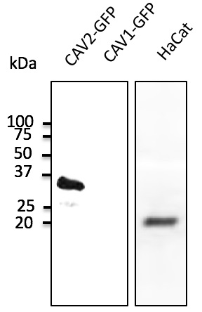
Figure 1. Western blot analysis of Caveolin-2/CAV2 using anti-Caveolin-2/CAV2 antibody (A01574). Electrophoresis was performed on a 5-20% SDS-PAGE gel at 70V (Stacking gel) / 90V (Resolving gel) for 2-3 hours. The sample well of each lane was loaded with 50ug of sample under reducing conditions. Lane 1: human Hela whole cell lysates, Lane 2: human A549 whole cell lysates, Lane 3: human U20S whole cell lysates, Lane 4: human K562 whole cell lysates, Lane 5: human HT1080 whole cell lysates, Lane 6: human CACO-2 whole cell lysates, Lane 7: human HEK293 whole cell lysates, Lane 8: monkey COS-7 whole cell lysates, Lane 9: rat skeletal muscle tissue lysates, Lane 10: rat heart tissue lysates, Lane 11: mouse skeletal muscle tissue lysates, Lane 12: mouse heart tissue lysates, Lane 13: mouse NIH/3T3 whole cell lysates. After Electrophoresis, proteins were transferred to a Nitrocellulose membrane at 150mA for 50-90 minutes. Blocked the membrane with 5% Non-fat Milk/ TBS for 1.5 hour at RT. The membrane was incubated with rabbit anti-Caveolin-2/CAV2 antigen affinity purified polyclonal antibody (Catalog # A01574) at 0.25 microg/mL overnight at 4°C, then washed with TBS-0.1%Tween 3 times with 5 minutes each and probed with a goat anti-rabbit IgG-HRP secondary antibody at a dilution of 1:10000 for 1.5 hour at RT. The signal is developed using an Enhanced Chemiluminescent detection (ECL) kit (Catalog # EK1002) with Tanon 5200 system. A specific band was detected for Caveolin-2/CAV2 at approximately 22KD. The expected band size for Caveolin-2/CAV2 is at 22KD.
. Caveolin-2/CAV2 was detected in paraffin-embedded section of human rectal cancer tissue. Heat mediated antigen retrieval was performed in EDTA buffer (pH8.0, epitope retrieval solution). The tissue section was blocked with 10% goat serum. The tissue section was then incubated with 1microg/ml rabbit anti-Caveolin-2/CAV2 Antibody (A01574) overnight at 4°C. Biotinylated goat anti-rabbit IgG was used as secondary antibody and incubated for 30 minutes at 37°C. The tissue section was developed using Strepavidin-Biotin-Complex (SABC) (Catalog # SA1022) with DAB as the chromogen.")
. Caveolin-2/CAV2 was detected in paraffin-embedded section of mouse lung tissue. Heat mediated antigen retrieval was performed in EDTA buffer (pH8.0, epitope retrieval solution). The tissue section was blocked with 10% goat serum. The tissue section was then incubated with 1microg/ml rabbit anti-Caveolin-2/CAV2 Antibody (A01574) overnight at 4°C. Biotinylated goat anti-rabbit IgG was used as secondary antibody and incubated for 30 minutes at 37°C. The tissue section was developed using Strepavidin-Biotin-Complex (SABC) (Catalog # SA1022) with DAB as the chromogen.")
. Caveolin-2/CAV2 was detected in paraffin-embedded section of rat lung tissue. Heat mediated antigen retrieval was performed in EDTA buffer (pH8.0, epitope retrieval solution). The tissue section was blocked with 10% goat serum. The tissue section was then incubated with 1microg/ml rabbit anti-Caveolin-2/CAV2 Antibody (A01574) overnight at 4°C. Biotinylated goat anti-rabbit IgG was used as secondary antibody and incubated for 30 minutes at 37°C. The tissue section was developed using Strepavidin-Biotin-Complex (SABC) (Catalog # SA1022) with DAB as the chromogen.")
. Caveolin-2/CAV2 was detected in paraffin-embedded section of rat lung tissue. Heat mediated antigen retrieval was performed in EDTA buffer (pH8.0, epitope retrieval solution). The tissue section was blocked with 10% goat serum. The tissue section was then incubated with 1microg/ml rabbit anti-Caveolin-2/CAV2 Antibody (A01574) overnight at 4°C. Biotinylated goat anti-rabbit IgG was used as secondary antibody and incubated for 30 minutes at 37°C. The tissue section was developed using Strepavidin-Biotin-Complex (SABC) (Catalog # SA1022) with DAB as the chromogen.")
. Caveolin-2/CAV2 was detected in immunocytochemical section of A549 cells. Enzyme antigen retrieval was performed using IHC enzyme antigen retrieval reagent (AR0022) for 15 mins. The cells were blocked with 10% goat serum. And then incubated with 5microg/mL rabbit anti-Caveolin-2/CAV2 Antibody (A01574) overnight at 4°C. DyLight®488 Conjugated Goat Anti-Rabbit IgG (BA1127) was used as secondary antibody at 1:100 dilution and incubated for 30 minutes at 37°C. The section was counterstained with DAPI. Visualize using a fluorescence microscope and filter sets appropriate for the label used.")
. Caveolin-2/CAV2 was detected in paraffin-embedded section of human intestinal cancer tissue. Heat mediated antigen retrieval was performed in EDTA buffer (pH8.0, epitope retrieval solution). The tissue section was blocked with 10% goat serum. The tissue section was then incubated with 5microg/mL rabbit anti-Caveolin-2/CAV2 Antibody (A01574) overnight at 4°C. DyLight®594 Conjugated Goat Anti-Rabbit IgG (BA1142) was used as secondary antibody at 1:100 dilution and incubated for 30 minutes at 37°C. The section was counterstained with DAPI. Visualize using a fluorescence microscope and filter sets appropriate for the label used.")
. Overlay histogram showing A549 cells stained with A01574 (Blue line). To facilitate intracellular staining, cells were fixed with 4% paraformaldehyde and permeabilized with permeabilization buffer. The cells were blocked with 10% normal goat serum. And then incubated with rabbit anti-Caveolin-2/CAV2 Antibody (A01574,1microg/1x106 cells) for 30 min at 20°C. DyLight®488 conjugated goat anti-rabbit IgG (BA1127, 5-10microg/1x106 cells) was used as secondary antibody for 30 minutes at 20°C. Isotype control antibody (Green line) was rabbit IgG (1microg/1x106) used under the same conditions. Unlabelled sample without incubation with primary antibody and secondary antibody (Red line) was used as a blank control.")
Figure 1. Western blot analysis of Caveolin-2/CAV2 using anti-Caveolin-2/CAV2 antibody (A01574). Electrophoresis was performed on a 5-20% SDS-PAGE gel at 70V (Stacking gel) / 90V (Resolving gel) for 2-3 hours. The sample well of each lane was loaded with 50ug of sample under reducing conditions. Lane 1: human Hela whole cell lysates, Lane 2: human A549 whole cell lysates, Lane 3: human U20S whole cell lysates, Lane 4: human K562 whole cell lysates, Lane 5: human HT1080 whole cell lysates, Lane 6: human CACO-2 whole cell lysates, Lane 7: human HEK293 whole cell lysates, Lane 8: monkey COS-7 whole cell lysates, Lane 9: rat skeletal muscle tissue lysates, Lane 10: rat heart tissue lysates, Lane 11: mouse skeletal muscle tissue lysates, Lane 12: mouse heart tissue lysates, Lane 13: mouse NIH/3T3 whole cell lysates. After Electrophoresis, proteins were transferred to a Nitrocellulose membrane at 150mA for 50-90 minutes. Blocked the membrane with 5% Non-fat Milk/ TBS for 1.5 hour at RT. The membrane was incubated with rabbit anti-Caveolin-2/CAV2 antigen affinity purified polyclonal antibody (Catalog # A01574) at 0.25 microg/mL overnight at 4°C, then washed with TBS-0.1%Tween 3 times with 5 minutes each and probed with a goat anti-rabbit IgG-HRP secondary antibody at a dilution of 1:10000 for 1.5 hour at RT. The signal is developed using an Enhanced Chemiluminescent detection (ECL) kit (Catalog # EK1002) with Tanon 5200 system. A specific band was detected for Caveolin-2/CAV2 at approximately 22KD. The expected band size for Caveolin-2/CAV2 is at 22KD.
Anti-Caveolin-2/CAV2 Antibody Picoband(r)
A01574
ApplicationsFlow Cytometry, ImmunoFluorescence, Western Blot, ELISA, ImmunoCytoChemistry, ImmunoHistoChemistry
Product group Antibodies
ReactivityHuman, Monkey, Mouse, Rat
TargetCAV2
Overview
- SupplierBoster Bio
- Product NameAnti-Caveolin-2/CAV2 Antibody Picoband(r)
- Delivery Days Customer9
- Application Supplier NoteTested Species: In-house tested species with positive results. Other applications have not been tested. Optimal dilutions should be determined by end users.
- ApplicationsFlow Cytometry, ImmunoFluorescence, Western Blot, ELISA, ImmunoCytoChemistry, ImmunoHistoChemistry
- CertificationResearch Use Only
- ClonalityPolyclonal
- Concentration500 ug/ml
- Gene ID858
- Target nameCAV2
- Target descriptioncaveolin 2
- Target synonymsCAV, caveolin-2, caveolae protein, 20-kD
- HostRabbit
- IsotypeIgG
- Protein IDP51636
- Protein NameCaveolin-2
- Scientific DescriptionBoster Bio Anti-Caveolin-2/CAV2 Antibody Picoband® catalog # A01574. Tested in ELISA, Flow Cytometry, IF, IHC, ICC, WB applications. This antibody reacts with Human, Monkey, Mouse, Rat. The brand Picoband indicates this is a premium antibody that guarantees superior quality, high affinity, and strong signals with minimal background in Western blot applications. Only our best-performing antibodies are designated as Picoband, ensuring unmatched performance.
- ReactivityHuman, Monkey, Mouse, Rat
- Storage Instruction-20°C,2°C to 8°C
- UNSPSC12352203
Related products
Product group Antibodies
Caveolin-2 Recombinant AntibodyBSM-61101R
ApplicationsImmunoFluorescence, ImmunoPrecipitation, Western Blot, ImmunoCytoChemistry, ImmunoHistoChemistry, ImmunoHistoChemistry Frozen, ImmunoHistoChemistry Paraffin
ReactivityHuman
TargetCAV2
- SizePrice
Product group Antibodies
CAV2 Polyclonal AntibodyCAC14710
ApplicationsImmunoFluorescence, Western Blot, ELISA, ImmunoHistoChemistry
TargetCAV2
- SizePrice
Product group Antibodies
Anti-Caveolin 2 Antibody107-10752
ApplicationsImmunoFluorescence, Western Blot, ImmunoCytoChemistry, ImmunoHistoChemistry, ImmunoHistoChemistry Paraffin
ReactivityHuman
TargetCAV2
- SizePrice
Product group Antibodies
Anti-CAV2 AntibodyA121612
ApplicationsImmunoFluorescence, Western Blot, ImmunoHistoChemistry, ImmunoHistoChemistry Frozen, ImmunoHistoChemistry Paraffin
ReactivityCanine, Human, Mouse
- SizePrice
Product group Antibodies

Caveolin 2 (Phospho-Tyr27) AntibodyABX012660
ApplicationsWestern Blot, ELISA
- SizePrice
Product group Antibodies
References
Caveolin 2 antibodyGTX108294
ApplicationsImmunoFluorescence, ImmunoPrecipitation, Western Blot, ImmunoCytoChemistry, ImmunoHistoChemistry, ImmunoHistoChemistry Paraffin
ReactivityHuman, Mouse, Rat
TargetCAV2
- SizePrice
Product group Antibodies

CAV2 / Caveolin 2 AntibodyLS-C760883
ApplicationsWestern Blot
ReactivityHuman, Monkey, Mouse, Rat
TargetCAV2
- SizePrice

Product group Antibodies
Anti-CAV2 AntibodyHPA044810
ApplicationsWestern Blot, ImmunoCytoChemistry, ImmunoHistoChemistry
ReactivityHuman
TargetCAV2
- SizePrice