


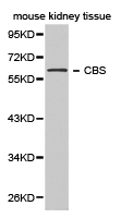
Figure 1. Western blot analysis of CBS using anti-CBS antibody (M00130-3). Electrophoresis was performed on a 5-20% SDS-PAGE gel at 70V (Stacking gel) / 90V (Resolving gel) for 2-3 hours. The sample well of each lane was loaded with 30 ug of sample under reducing conditions. Lane 1: human Hela whole cell lysates, Lane 2: human 293T whole cell lysates, Lane 3: human K562 whole cell lysates, Lane 4: rat liver tissue lysates, Lane 5: rat pancreas tissue lysates, Lane 6: mouse liver tissue lysates, Lane 7: mouse pancreas tissue lysates. After electrophoresis, proteins were transferred to a nitrocellulose membrane at 150 mA for 50-90 minutes. Blocked the membrane with 5% non-fat milk/TBS for 1.5 hour at RT. The membrane was incubated with mouse anti-CBS antigen affinity purified monoclonal antibody (Catalog # M00130-3) at 0.5 microg/mL overnight at 4°C, then washed with TBS-0.1%Tween 3 times with 5 minutes each and probed with a goat anti-mouse IgG-HRP secondary antibody at a dilution of 1:10000 for 1.5 hour at RT. The signal is developed using an Enhanced Chemiluminescent detection (ECL) kit (Catalog # EK1001) with Tanon 5200 system. A specific band was detected for CBS at approximately 61 kDa. The expected band size for CBS is at 61 kDa.
. Overlay histogram showing MCF-7 cells stained with M00130-3 (Blue line). To facilitate intracellular staining, cells were fixed with 4% paraformaldehyde and permeabilized with permeabilization buffer. The cells were blocked with 10% normal goat serum. And then incubated with mouse anti-CBS Antibody (M00130-3, 1 microg/1x106 cells) for 30 min at 20°C. DyLight®488 conjugated goat anti-mouse IgG (BA1126, 5-10 microg/1x106 cells) was used as secondary antibody for 30 minutes at 20°C. Isotype control antibody (Green line) was mouse IgG (1 microg/1x106) used under the same conditions. Unlabelled sample without incubation with primary antibody and secondary antibody (Red line) was used as a blank control.")
Figure 1. Western blot analysis of CBS using anti-CBS antibody (M00130-3). Electrophoresis was performed on a 5-20% SDS-PAGE gel at 70V (Stacking gel) / 90V (Resolving gel) for 2-3 hours. The sample well of each lane was loaded with 30 ug of sample under reducing conditions. Lane 1: human Hela whole cell lysates, Lane 2: human 293T whole cell lysates, Lane 3: human K562 whole cell lysates, Lane 4: rat liver tissue lysates, Lane 5: rat pancreas tissue lysates, Lane 6: mouse liver tissue lysates, Lane 7: mouse pancreas tissue lysates. After electrophoresis, proteins were transferred to a nitrocellulose membrane at 150 mA for 50-90 minutes. Blocked the membrane with 5% non-fat milk/TBS for 1.5 hour at RT. The membrane was incubated with mouse anti-CBS antigen affinity purified monoclonal antibody (Catalog # M00130-3) at 0.5 microg/mL overnight at 4°C, then washed with TBS-0.1%Tween 3 times with 5 minutes each and probed with a goat anti-mouse IgG-HRP secondary antibody at a dilution of 1:10000 for 1.5 hour at RT. The signal is developed using an Enhanced Chemiluminescent detection (ECL) kit (Catalog # EK1001) with Tanon 5200 system. A specific band was detected for CBS at approximately 61 kDa. The expected band size for CBS is at 61 kDa.
Anti-CBS Antibody Picoband(r) (monoclonal, 5B5D7)
M00130-3-BIOTIN
ApplicationsFlow Cytometry, Western Blot
Product group Antibodies
ReactivityHuman, Mouse, Rat
TargetCBS
Overview
- SupplierBoster Bio
- Product NameAnti-CBS Antibody Picoband(r) (monoclonal, 5B5D7)
- Delivery Days Customer9
- ApplicationsFlow Cytometry, Western Blot
- CertificationResearch Use Only
- ClonalityMonoclonal
- Clone ID5B5D7
- Concentration500 ug/ml
- ConjugateBiotin
- Gene ID875
- Target nameCBS
- Target descriptioncystathionine beta-synthase
- Target synonymsCBSL, HIP4, cystathionine beta-synthase, Cystathionine beta-synthase-like protein, beta-thionase, methylcysteine synthase, serine sulfhydrase
- HostMouse
- IsotypeIgG1
- Protein IDP35520
- Protein NameCystathionine beta-synthase
- Scientific DescriptionBoster Bio Anti-CBS Antibody Picoband® (monoclonal, 5B5D7) catalog # M00130-3. Tested in Flow Cytometry, WB applications. This antibody reacts with Human, Mouse, Rat. The brand Picoband indicates this is a premium antibody that guarantees superior quality, high affinity, and strong signals with minimal background in Western blot applications. Only our best-performing antibodies are designated as Picoband, ensuring unmatched performance.
- ReactivityHuman, Mouse, Rat
- Storage Instruction-20°C,2°C to 8°C
- UNSPSC12352203
Related products
Product group Antibodies
Anti-CBS Antibody144-01427
ApplicationsImmunoFluorescence, Western Blot, ImmunoHistoChemistry
ReactivityHuman, Mouse
TargetCBS
- SizePrice
Product group Antibodies
Cbs Polyclonal AntibodyCAC11162
ApplicationsImmunoFluorescence, ImmunoPrecipitation, Western Blot, ELISA, ImmunoHistoChemistry
ReactivityMouse
TargetCBS
- SizePrice
Product group Antibodies
Anti-CBS AntibodyA29799
ApplicationsImmunoFluorescence, Western Blot, ImmunoHistoChemistry
ReactivityHuman, Mouse, Rat
- SizePrice

Product group Antibodies
Anti-CBS Antibody Picoband(r)A00130-3-CARRIER-FREE
ApplicationsFlow Cytometry, ImmunoFluorescence, Western Blot, ELISA, ImmunoCytoChemistry
ReactivityHuman, Mouse, Rat
TargetCBS
- SizePrice
Product group Antibodies
CBS AntibodyCSB-PA12719A0RB
ApplicationsImmunoFluorescence, ImmunoPrecipitation, Western Blot, ELISA, ImmunoHistoChemistry
ReactivityHuman, Mouse
TargetCBS
- SizePrice
Product group Antibodies
References
CBS antibodyGTX113400
ApplicationsImmunoFluorescence, ImmunoPrecipitation, Western Blot, ImmunoCytoChemistry, ImmunoHistoChemistry, ImmunoHistoChemistry Paraffin
ReactivityHuman, Mouse, Rat
TargetCBS
- SizePrice
Product group Antibodies
CBS Recombinant Antibody, AbBy Fluor-405 ConjugatedBSM-61957R-BF405
ApplicationsImmunoFluorescence, Western Blot, ImmunoCytoChemistry
ReactivityHuman
TargetCBS
- SizePrice

Product group Antibodies
Anti-CBS AntibodyHPA001223
ApplicationsImmunoCytoChemistry, ImmunoHistoChemistry
ReactivityHuman
TargetCBS
- SizePrice