


Immunofluorescent staining of human cell line HAP1 shows localization to plasma membrane.
Immunofluorescent staining of human cell line HAP1 shows localization to plasma membrane.
Anti-CCKBR Antibody
HPA079548
ApplicationsImmunoCytoChemistry
Product group Antibodies
ReactivityHuman
TargetCCKBR
Overview
- SupplierAtlas Antibodies
- Product NameAnti-CCKBR Antibody
- Delivery Days Customer12
- ApplicationsImmunoCytoChemistry
- CertificationResearch Use Only
- ClonalityPolyclonal
- ConjugateUnconjugated
- Gene ID887
- Target nameCCKBR
- Target descriptioncholecystokinin B receptor
- Target synonymsCCK-2R, CCK-B, CCK2R, GASR, gastrin/cholecystokinin type B receptor, CCK-B receptor, CCK2 receptor, cholecystokinin type 2 receptor, cholecystokinin-2 receptor, gastrin receptor
- HostRabbit
- IsotypeIgG
- Protein IDP32239
- Protein NameGastrin/cholecystokinin type B receptor
- Scientific DescriptionRecombinant Protein Epitope Signature Tag (PrEST) antigen sequence
- ReactivityHuman
- Storage Instruction-20°C,2°C to 8°C
- UNSPSC41116161
MSDS
Related products
Product group Antibodies
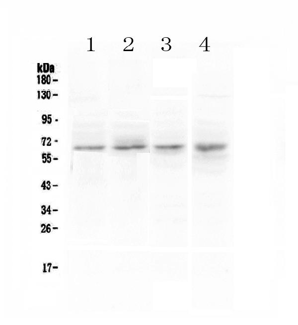
CCKBR AntibodyCSB-PA001401
ApplicationsWestern Blot, ELISA
ReactivityHuman, Monkey, Mouse, Rat
TargetCCKBR
- SizePrice
Product group Antibodies
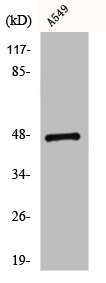
Anti-CCKBR Antibody Picoband(r)A01677-1-CARRIER-FREE
ApplicationsFlow Cytometry, Western Blot, ImmunoCytoChemistry, ImmunoHistoChemistry
ReactivityHuman, Mouse, Rat
TargetCCKBR
- SizePrice
Product group Antibodies
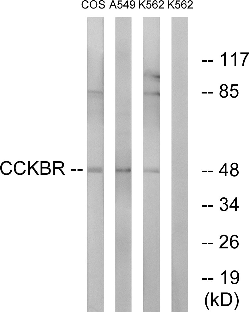
Anti-CCKBR AntibodyA97658
ApplicationsWestern Blot, ELISA
ReactivityHuman, Mouse, Rat
- SizePrice
Product group Antibodies

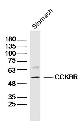
CCKBR / Cckb AntibodyLS-C749569
ApplicationsWestern Blot
ReactivityHuman, Mouse, Rat
TargetCCKBR
- SizePrice
Product group Antibodies
References

Goat anti-CCKBREB06767
ApplicationsWestern Blot, ELISA, ImmunoHistoChemistry
ReactivityBovine, Human, Mouse, Rat
TargetCCKBR
- SizePrice
Product group Antibodies
References
CCKBR Polyclonal AntibodyBS-1777R
ApplicationsImmunoFluorescence, Western Blot, ELISA, ImmunoCytoChemistry, ImmunoHistoChemistry, ImmunoHistoChemistry Frozen, ImmunoHistoChemistry Paraffin
ReactivityBovine, Human, Mouse, Rabbit, Rat
TargetCCKBR
- SizePrice