

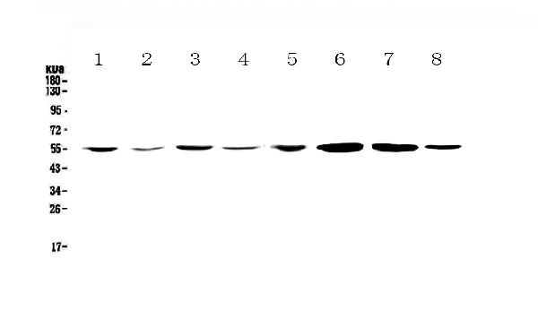
Figure 1. Western blot analysis of CCR3 using anti-CCR3 antibody (A01748-1). Electrophoresis was performed on a 5-20% SDS-PAGE gel at 70V (Stacking gel) / 90V (Resolving gel) for 2-3 hours. The sample well of each lane was loaded with 50ug of sample under reducing conditions. Lane 1: human Jurkat whole cell lysates, Lane 2: human HepG2 whole cell lysates, Lane 3: human MCF-7 whole cell lysates, Lane 4: human U-87MG whole cell lysates, Lane 5: human CCRF-CEM whole cell lysates, Lane 6: rat brain tissue lysates, Lane 7: mouse brain tissue lysates, Lane 8: mouse testis tissue lysates. After Electrophoresis, proteins were transferred to a Nitrocellulose membrane at 150mA for 50-90 minutes. Blocked the membrane with 5% Non-fat Milk/ TBS for 1.5 hour at RT. The membrane was incubated with rabbit anti-CCR3 antigen affinity purified polyclonal antibody (Catalog # A01748-1) at 0.5 microg/mL overnight at 4°C, then washed with TBS-0.1%Tween 3 times with 5 minutes each and probed with a goat anti-rabbit IgG-HRP secondary antibody at a dilution of 1:10000 for 1.5 hour at RT. The signal is developed using an Enhanced Chemiluminescent detection (ECL) kit (Catalog # EK1002) with Tanon 5200 system. A specific band was detected for CCR3 at approximately 55KD. The expected band size for CCR3 is at 41KD.
. Overlay histogram showing RAW264.7 cells stained with A01748-1 (Blue line). The cells were fixed with 4% paraformaldehyde and blocked with 10% normal goat serum. And then incubated with rabbit anti-CCR3 Antibody (A01748-1,1microg/1x106 cells) for 30 min at 20°C. DyLight®488 conjugated goat anti-rabbit IgG (BA1127, 5-10microg/1x106 cells) was used as secondary antibody for 30 minutes at 20°C. Isotype control antibody (Green line) was rabbit IgG (1microg/1x106) used under the same conditions. Unlabelled sample without incubation with primary antibody and secondary antibody (Red line) was used as a blank control.")
Figure 1. Western blot analysis of CCR3 using anti-CCR3 antibody (A01748-1). Electrophoresis was performed on a 5-20% SDS-PAGE gel at 70V (Stacking gel) / 90V (Resolving gel) for 2-3 hours. The sample well of each lane was loaded with 50ug of sample under reducing conditions. Lane 1: human Jurkat whole cell lysates, Lane 2: human HepG2 whole cell lysates, Lane 3: human MCF-7 whole cell lysates, Lane 4: human U-87MG whole cell lysates, Lane 5: human CCRF-CEM whole cell lysates, Lane 6: rat brain tissue lysates, Lane 7: mouse brain tissue lysates, Lane 8: mouse testis tissue lysates. After Electrophoresis, proteins were transferred to a Nitrocellulose membrane at 150mA for 50-90 minutes. Blocked the membrane with 5% Non-fat Milk/ TBS for 1.5 hour at RT. The membrane was incubated with rabbit anti-CCR3 antigen affinity purified polyclonal antibody (Catalog # A01748-1) at 0.5 microg/mL overnight at 4°C, then washed with TBS-0.1%Tween 3 times with 5 minutes each and probed with a goat anti-rabbit IgG-HRP secondary antibody at a dilution of 1:10000 for 1.5 hour at RT. The signal is developed using an Enhanced Chemiluminescent detection (ECL) kit (Catalog # EK1002) with Tanon 5200 system. A specific band was detected for CCR3 at approximately 55KD. The expected band size for CCR3 is at 41KD.
Anti-CCR3 Antibody Picoband(r)
A01748-1-CARRIER-FREE
ApplicationsFlow Cytometry, Western Blot
Product group Antibodies
ReactivityHuman, Mouse, Rat
TargetCCR3
Overview
- SupplierBoster Bio
- Product NameAnti-CCR3 Antibody Picoband(r)
- Delivery Days Customer9
- ApplicationsFlow Cytometry, Western Blot
- CertificationResearch Use Only
- ClonalityPolyclonal
- Concentration500 ug/ml
- Gene ID1232
- Target nameCCR3
- Target descriptionC-C motif chemokine receptor 3
- Target synonymsC C CKR3, CC-CKR-3, CD193, CKR 3, CKR3, CMKBR3, C-C chemokine receptor type 3, C-C CKR-3, CC chemokine receptor 3, CCR-3, b-chemokine receptor, chemokine (C-C motif) receptor 3, eosinophil CC chemokine receptor 3, eosinophil eotaxin receptor
- HostRabbit
- IsotypeIgG
- Protein IDP51677
- Protein NameC-C chemokine receptor type 3
- Scientific DescriptionBoster Bio Anti-CCR3 Antibody Picoband® catalog # A01748-1. Tested in Flow Cytometry, WB applications. This antibody reacts with Human, Mouse, Rat. The brand Picoband indicates this is a premium antibody that guarantees superior quality, high affinity, and strong signals with minimal background in Western blot applications. Only our best-performing antibodies are designated as Picoband, ensuring unmatched performance.
- ReactivityHuman, Mouse, Rat
- Storage Instruction-20°C,2°C to 8°C
- UNSPSC12352203
Related products
Product group Antibodies

Anti-CCR3 (CD193) [5E8-G9-B4]Ab00223-23.0
ApplicationsFlow Cytometry
ReactivityHuman
TargetCCR3
- SizePrice
Product group Antibodies
Anti-Mouse CCR3 (aa5-28) Antibody119-16951
ApplicationsImmunoHistoChemistry, ImmunoHistoChemistry Paraffin
ReactivityHuman, Mouse
TargetCCR3
- SizePrice
Product group Antibodies
Anti-CCR3 AntibodyA101528
ApplicationsWestern Blot, ELISA
ReactivityHuman
- SizePrice
Product group Antibodies

CCR3 AntibodyLS-C677420
ApplicationsImmunoFluorescence, Western Blot, ELISA, ImmunoHistoChemistry, ImmunoHistoChemistry Paraffin
ReactivityHuman
TargetCCR3
- SizePrice
Product group Antibodies
CCR3 Recombinant AntibodyBSM-61221R
ApplicationsWestern Blot
TargetCCR3
- SizePrice
Product group Antibodies
CCR3 Polyclonal AntibodyCAC14934
ApplicationsImmunoFluorescence, Western Blot, ELISA, ImmunoHistoChemistry
ReactivityMouse
TargetCCR3
- SizePrice
Product group Antibodies
CCR3 AntibodyCSB-PA004842LA01HU
ApplicationsImmunoFluorescence, Western Blot, ELISA, ImmunoHistoChemistry
ReactivityHuman, Mouse
TargetCCR3
- SizePrice
![FACS analysis of human peripheral blood using GTX00536 CCR3 antibody [500000000].](https://www.genetex.com/upload/website/prouct_img/normal/GTX00536/GTX00536_20191025_AP_006_79_w_23053121_552.webp)
Product group Antibodies
CCR3 antibody [5E8]GTX00536
ApplicationsFlow Cytometry
ReactivityHuman
TargetCCR3
- SizePrice

Product group Antibodies
Anti-CCR3 AntibodyHPA069514
ApplicationsImmunoCytoChemistry
ReactivityHuman
TargetCCR3
- SizePrice