

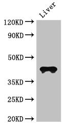
Western Blot Positive WB detected in: Mouse liver tissue All lanes: CCR3 antibody at 3ug/ml Secondary Goat polyclonal to rabbit IgG at 1/50000 dilution predicted band size: 42, 44 kDa observed band size: 42 kDa

")
Western Blot Positive WB detected in: Mouse liver tissue All lanes: CCR3 antibody at 3ug/ml Secondary Goat polyclonal to rabbit IgG at 1/50000 dilution predicted band size: 42, 44 kDa observed band size: 42 kDa
CCR3 Antibody
CSB-PA004842LA01HU
ApplicationsImmunoFluorescence, Western Blot, ELISA, ImmunoHistoChemistry
Product group Antibodies
ReactivityHuman, Mouse
TargetCCR3
Overview
- SupplierCusabio
- Product NameCCR3 Antibody
- Delivery Days Customer20
- ApplicationsImmunoFluorescence, Western Blot, ELISA, ImmunoHistoChemistry
- CertificationResearch Use Only
- ClonalityPolyclonal
- ConjugateUnconjugated
- Gene ID1232
- Target nameCCR3
- Target descriptionC-C motif chemokine receptor 3
- Target synonymsC C CKR3, CC-CKR-3, CD193, CKR 3, CKR3, CMKBR3, C-C chemokine receptor type 3, C-C CKR-3, CC chemokine receptor 3, CCR-3, b-chemokine receptor, chemokine (C-C motif) receptor 3, eosinophil CC chemokine receptor 3, eosinophil eotaxin receptor
- HostRabbit
- IsotypeIgG
- Protein IDP51677
- Protein NameC-C chemokine receptor type 3
- Scientific DescriptionReceptor for a C-C type chemokine. Binds to eotaxin, eotaxin-3, MCP-3, MCP-4, RANTES and MIP-1 delta. Subsequently transduces a signal by increasing the intracellular calcium ions level. Alternative coreceptor with CD4 for HIV-1 infection.
- ReactivityHuman, Mouse
- Storage Instruction-20°C or -80°C
- UNSPSC41116161
Related products
Product group Antibodies

Anti-CCR3 (CD193) [5E8-G9-B4]Ab00223-23.0
ApplicationsFlow Cytometry
ReactivityHuman
TargetCCR3
- SizePrice
Product group Antibodies
Anti-Mouse CCR3 (aa5-28) Antibody119-16951
ApplicationsImmunoHistoChemistry, ImmunoHistoChemistry Paraffin
ReactivityHuman, Mouse
TargetCCR3
- SizePrice
Product group Antibodies
Anti-CCR3 AntibodyA101528
ApplicationsWestern Blot, ELISA
ReactivityHuman
- SizePrice
Product group Antibodies

CCR3 AntibodyLS-C677420
ApplicationsImmunoFluorescence, Western Blot, ELISA, ImmunoHistoChemistry, ImmunoHistoChemistry Paraffin
ReactivityHuman
TargetCCR3
- SizePrice
Product group Antibodies
Anti-CCR3 Antibody Picoband(r)A01748-1-CARRIER-FREE
ApplicationsFlow Cytometry, Western Blot
ReactivityHuman, Mouse, Rat
TargetCCR3
- SizePrice
Product group Antibodies
CCR3 Recombinant AntibodyBSM-61221R
ApplicationsWestern Blot
TargetCCR3
- SizePrice
Product group Antibodies
CCR3 Polyclonal AntibodyCAC14934
ApplicationsImmunoFluorescence, Western Blot, ELISA, ImmunoHistoChemistry
ReactivityMouse
TargetCCR3
- SizePrice
![FACS analysis of human peripheral blood using GTX00536 CCR3 antibody [500000000].](https://www.genetex.com/upload/website/prouct_img/normal/GTX00536/GTX00536_20191025_AP_006_79_w_23053121_552.webp)
Product group Antibodies
CCR3 antibody [5E8]GTX00536
ApplicationsFlow Cytometry
ReactivityHuman
TargetCCR3
- SizePrice

Product group Antibodies
Anti-CCR3 AntibodyHPA069514
ApplicationsImmunoCytoChemistry
ReactivityHuman
TargetCCR3
- SizePrice