

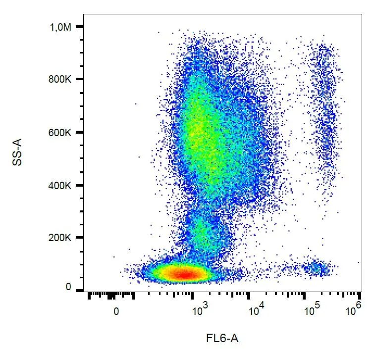
FACS analysis of human peripheral blood using GTX00536 CCR3 antibody [500000000].
FACS analysis of human peripheral blood using GTX00536 CCR3 antibody [500000000].
CCR3 antibody [5E8]
GTX00536
ApplicationsFlow Cytometry
Product group Antibodies
ReactivityHuman
TargetCCR3
Overview
- SupplierGeneTex
- Product NameCCR3 antibody [5E8]
- Delivery Days Customer9
- ApplicationsFlow Cytometry
- CertificationResearch Use Only
- ClonalityMonoclonal
- Concentration1 mg/ml
- ConjugateUnconjugated
- Gene ID1232
- Target nameCCR3
- Target descriptionC-C motif chemokine receptor 3
- Target synonymsC C CKR3, CC-CKR-3, CD193, CKR 3, CKR3, CMKBR3, C-C chemokine receptor type 3, C-C CKR-3, CC chemokine receptor 3, CCR-3, b-chemokine receptor, chemokine (C-C motif) receptor 3, eosinophil CC chemokine receptor 3, eosinophil eotaxin receptor
- HostMouse
- IsotypeIgG2b
- Protein IDP51677
- Protein NameC-C chemokine receptor type 3
- Scientific DescriptionThe protein encoded by this gene is a receptor for C-C type chemokines. It belongs to family 1 of the G protein-coupled receptors. This receptor binds and responds to a variety of chemokines, including eotaxin (CCL11), eotaxin-3 (CCL26), MCP-3 (CCL7), MCP-4 (CCL13), and RANTES (CCL5). It is highly expressed in eosinophils and basophils, and is also detected in TH1 and TH2 cells, as well as in airway epithelial cells. This receptor may contribute to the accumulation and activation of eosinophils and other inflammatory cells in the allergic airway. It is also known to be an entry co-receptor for HIV-1. This gene and seven other chemokine receptor genes form a chemokine receptor gene cluster on the chromosomal region 3p21. Alternatively spliced transcript variants have been described. [provided by RefSeq, Sep 2009]
- ReactivityHuman
- Storage Instruction2°C to 8°C
- UNSPSC41116161
Datasheet
Related products
Product group Antibodies

Anti-CCR3 (CD193) [5E8-G9-B4]Ab00223-23.0
ApplicationsFlow Cytometry
ReactivityHuman
TargetCCR3
- SizePrice
Product group Antibodies
Anti-Mouse CCR3 (aa5-28) Antibody119-16951
ApplicationsImmunoHistoChemistry, ImmunoHistoChemistry Paraffin
ReactivityHuman, Mouse
TargetCCR3
- SizePrice
Product group Antibodies
Anti-CCR3 AntibodyA101528
ApplicationsWestern Blot, ELISA
ReactivityHuman
- SizePrice
Product group Antibodies

CCR3 AntibodyLS-C677420
ApplicationsImmunoFluorescence, Western Blot, ELISA, ImmunoHistoChemistry, ImmunoHistoChemistry Paraffin
ReactivityHuman
TargetCCR3
- SizePrice
Product group Antibodies
Anti-CCR3 Antibody Picoband(r)A01748-1-CARRIER-FREE
ApplicationsFlow Cytometry, Western Blot
ReactivityHuman, Mouse, Rat
TargetCCR3
- SizePrice
Product group Antibodies
CCR3 Recombinant AntibodyBSM-61221R
ApplicationsWestern Blot
TargetCCR3
- SizePrice
Product group Antibodies
CCR3 Polyclonal AntibodyCAC14934
ApplicationsImmunoFluorescence, Western Blot, ELISA, ImmunoHistoChemistry
ReactivityMouse
TargetCCR3
- SizePrice
Product group Antibodies
CCR3 AntibodyCSB-PA004842LA01HU
ApplicationsImmunoFluorescence, Western Blot, ELISA, ImmunoHistoChemistry
ReactivityHuman, Mouse
TargetCCR3
- SizePrice

Product group Antibodies
CCR3 antibodyGTX21667
ApplicationsImmunoFluorescence, Western Blot, ELISA, ImmunoCytoChemistry, ImmunoHistoChemistry
ReactivityHuman
TargetCCR3
- SizePrice

Product group Antibodies
CCR3 antibodyGTX31254
ApplicationsWestern Blot, ELISA, ImmunoHistoChemistry, ImmunoHistoChemistry Paraffin
ReactivityHuman, Mouse, Rat
TargetCCR3
- SizePrice