

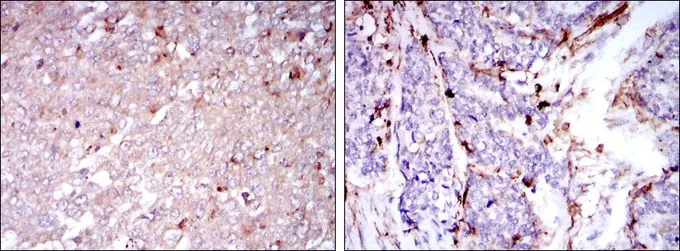
IHC-P analysis of human breast cancer tissues (left) and human esophageal cancer tissues (right) using GTX60471 CD133 antibody [3F10]
![ELISA analysis of antigen using GTX60471 CD133 antibody [3F10].
Red : Control antigen 100ng
Purple : Antigen 10ng
Green : Antigen 50ng
Blue : Antigen 100ng](https://www.genetex.com/upload/website/prouct_img/normal/GTX60471/GTX60471_20170912_ELISA_w_23061123_107.webp "ELISA analysis of antigen using GTX60471 CD133 antibody [3F10].
Red : Control antigen 100ng
Purple : Antigen 10ng
Green : Antigen 50ng
Blue : Antigen 100ng")
![FACS analysis of HeLa cells using GTX60471 CD133 antibody [3F10]. Blue : CD133 Red : negative control](https://www.genetex.com/upload/website/prouct_img/normal/GTX60471/GTX60471_20170912_FACS_w_23061123_489.webp "FACS analysis of HeLa cells using GTX60471 CD133 antibody [3F10]. Blue : CD133 Red : negative control")
IHC-P analysis of human breast cancer tissues (left) and human esophageal cancer tissues (right) using GTX60471 CD133 antibody [3F10]
CD133 antibody [3F10]
GTX60471
ApplicationsFlow Cytometry, ELISA, ImmunoHistoChemistry, ImmunoHistoChemistry Paraffin
Product group Antibodies
ReactivityHuman
TargetPROM1
Overview
- SupplierGeneTex
- Product NameCD133 antibody [3F10]
- Delivery Days Customer9
- Application Supplier NoteIHC-P: 1/200 - 1/1000. FACS: 1/200 - 1/400. ELISA: 1/10000. *Optimal dilutions/concentrations should be determined by the researcher.Not tested in other applications.
- ApplicationsFlow Cytometry, ELISA, ImmunoHistoChemistry, ImmunoHistoChemistry Paraffin
- CertificationResearch Use Only
- ClonalityMonoclonal
- Clone ID3F10
- ConjugateUnconjugated
- Gene ID8842
- Target namePROM1
- Target descriptionprominin 1
- Target synonymsAC133, CD133, CORD12, MCDR2, MSTP061, PROML1, RP41, STGD4, prominin-1, AC133 antigen, Retinitis pigmentosa 41, Cone-rod dystrophy 12, antigen AC133, hProminin, hematopoietic stem cell antigen, prominin-like protein 1
- HostMouse
- IsotypeIgG1
- Protein IDO43490
- Protein NameProminin-1
- Scientific DescriptionThis gene encodes a pentaspan transmembrane glycoprotein. The protein localizes to membrane protrusions and is often expressed on adult stem cells, where it is thought to function in maintaining stem cell properties by suppressing differentiation. Mutations in this gene have been shown to result in retinitis pigmentosa and Stargardt disease. Expression of this gene is also associated with several types of cancer. This gene is expressed from at least five alternative promoters that are expressed in a tissue-dependent manner. Multiple transcript variants encoding different isoforms have been found for this gene. [provided by RefSeq, Mar 2009]
- ReactivityHuman
- Storage Instruction-20°C or -80°C,2°C to 8°C
- UNSPSC12352203
References
- Kim J, Shin K, Lee SH, et al. Slug and CD133 expression are associated with peritoneal carcinomatosis and survival in gastric cancer. J Gastrointest Oncol. 2021,12(4):1326-1337. doi: 10.21037/jgo-21-123Read this paper
- Luna ECM, Bezerra TMM, Barros Silva PG, et al. CD133 Role in Oral Carcinogenesis. Asian Pac J Cancer Prev. 2020,21(9):2501-2506. doi: 10.31557/APJCP.2020.21.9.2501Read this paper
Datasheet
Related products
Product group Antibodies

Anti-CD133 [CD133-scFv-1]AB01481-10.0-BT
ApplicationsFlow Cytometry, Western Blot, ELISA
ReactivityHuman
TargetPROM1
- SizePrice
Product group Antibodies
ApplicationsFlow Cytometry
ReactivityHuman
TargetPROM1
- SizePrice

Product group Antibodies
Anti-PROM1 Antibody Picoband(r)A01767-3-CARRIER-FREE
ApplicationsFlow Cytometry, ImmunoFluorescence, Western Blot, ELISA, ImmunoCytoChemistry
ReactivityHuman
TargetPROM1
- SizePrice
![Various whole cell extracts (30 μg) were separated by 5% SDS-PAGE, and the membrane was blotted with CD133 antibody [C1C2], Internal (GTX100567) diluted at 1:500. The HRP-conjugated anti-rabbit IgG antibody (GTX213110-01) was used to detect the primary antibody.](https://www.genetex.com/upload/website/prouct_img/normal/GTX100567/GTX100567_39471_20190530_WB_w_23060100_319.webp)
Product group Antibodies
References
CD133 antibody [C1C2], InternalGTX100567
ApplicationsFlow Cytometry, ImmunoFluorescence, Western Blot, ImmunoCytoChemistry, ImmunoHistoChemistry, ImmunoHistoChemistry Paraffin
ReactivityHuman
TargetPROM1
- SizePrice
![Various whole cell extracts (30 μg) were separated by 5% SDS-PAGE, and the membrane was blotted with CD133 antibody [N1C1] (GTX102109) diluted at 1:500. The HRP-conjugated anti-rabbit IgG antibody (GTX213110-01) was used to detect the primary antibody, and the signal was developed with Trident ECL plus-Enhanced.](https://www.genetex.com/upload/website/prouct_img/normal/GTX102109/GTX102109_39549_20190530_WB_w_23060100_233.webp)
Product group Antibodies
References
CD133 antibody [N1C1]GTX102109
ApplicationsWestern Blot, ImmunoHistoChemistry, ImmunoHistoChemistry Paraffin
ReactivityHuman
TargetPROM1
- SizePrice
![Various whole cell extracts (30 μg) were separated by 7.5% SDS-PAGE, and the membrane was blotted with CD133 antibody [HL1271] (GTX636673) diluted at 1:1000. The HRP-conjugated anti-rabbit IgG antibody (GTX213110-01) was used to detect the primary antibody.](https://www.genetex.com/upload/website/prouct_img/normal/GTX636673/GTX636673_44599_20220225_WB_w_23061202_503.webp)
Product group Antibodies
CD133 antibody [HL1271]GTX636673
ApplicationsWestern Blot
ReactivityHuman
TargetPROM1
- SizePrice
![CD133 antibody [HL1631] (GTX637124) detects CD133 protein by flow cytometry analysis. Sample: NT2D1 cell. Gray: Rabbit IgG Isotype control. Red: CD133 antibody [HL1631] (GTX637124) dilution: 1:50. Acquisition of 20,000 events were collected for FACS analysis. *The competitor (clone [AC133]) is not affiliated with GeneTex and does not endorse this product.](https://www.genetex.com/upload/website/prouct_img/normal/GTX637124/GTX637124_T-44739_20220722_FACS_22081000_264.webp)
Product group Antibodies
CD133 antibody [HL1631]GTX637124
ApplicationsFlow Cytometry
ReactivityHuman
TargetPROM1
- SizePrice
Product group Antibodies
Prom1 Polyclonal AntibodyCAC08759
ApplicationsWestern Blot, ELISA, ImmunoHistoChemistry
TargetPROM1
- SizePrice