


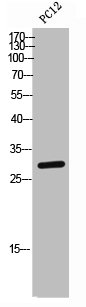
Figure 1. Western blot analysis of CD134 using anti-CD134 antibody (PB9898). Electrophoresis was performed on a 5-20% SDS-PAGE gel at 70V (Stacking gel) / 90V (Resolving gel) for 2-3 hours. The sample well of each lane was loaded with 50ug of sample under reducing conditions. Lane 1: SW620 whole cell lysates, Lane 2: 22RV1 whole cell lysates. After Electrophoresis, proteins were transferred to a Nitrocellulose membrane at 150mA for 50-90 minutes. Blocked the membrane with 5% Non-fat Milk/ TBS for 1.5 hour at RT. The membrane was incubated with rabbit anti-CD134 antigen affinity purified polyclonal antibody (Catalog # PB9898) at 0.5 microg/mL overnight at 4°C, then washed with TBS-0.1%Tween 3 times with 5 minutes each and probed with a goat anti-rabbit IgG-HRP secondary antibody at a dilution of 1:10000 for 1.5 hour at RT. The signal is developed using an Enhanced Chemiluminescent detection (ECL) kit (Catalog # EK1002) with Tanon 5200 system. A specific band was detected for CD134 at approximately 50KD. The expected band size for CD134 is at 50KD.
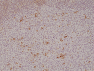
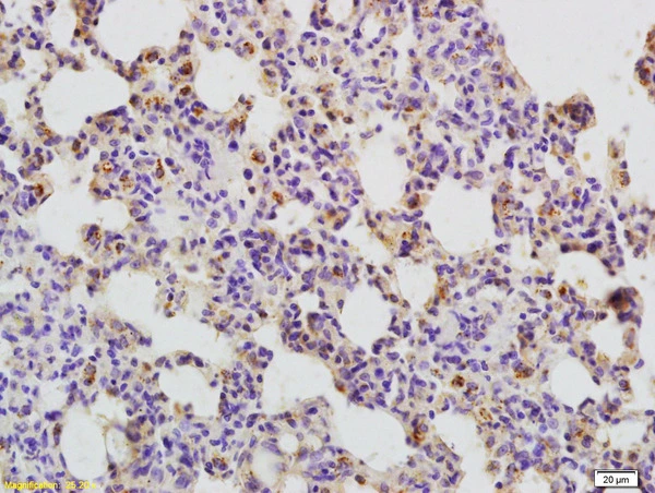
. CD134 was detected in paraffin-embedded section of human tonsil tissues. Heat mediated antigen retrieval was performed in citrate buffer (pH6, epitope retrieval solution) for 20 mins. The tissue section was blocked with 10% goat serum. The tissue section was then incubated with 1microg/ml rabbit anti-CD134 Antibody (PB9898) overnight at 4°C. Biotinylated goat anti-rabbit IgG was used as secondary antibody and incubated for 30 minutes at 37°C. The tissue section was developed using Strepavidin-Biotin-Complex (SABC)(Catalog # SA1022) with DAB as the chromogen.")
. Overlay histogram showing H-PBMC cells stained with PB9898 (Blue line). The cells were fixed with 4% paraformaldehyde and blocked with 10% normal goat serum. And then incubated with rabbit anti-CD134 Antibody (PB9898,1microg/1x106 cells) for 30 min at 20°C. DyLight®488 conjugated goat anti-rabbit IgG (BA1127, 5-10microg/1x106 cells) was used as secondary antibody for 30 minutes at 20°C. Isotype control antibody (Green line) was rabbit IgG (1microg/1x106) used under the same conditions. Unlabelled sample without incubation with primary antibody and secondary antibody (Red line) was used as a blank control.")
Figure 1. Western blot analysis of CD134 using anti-CD134 antibody (PB9898). Electrophoresis was performed on a 5-20% SDS-PAGE gel at 70V (Stacking gel) / 90V (Resolving gel) for 2-3 hours. The sample well of each lane was loaded with 50ug of sample under reducing conditions. Lane 1: SW620 whole cell lysates, Lane 2: 22RV1 whole cell lysates. After Electrophoresis, proteins were transferred to a Nitrocellulose membrane at 150mA for 50-90 minutes. Blocked the membrane with 5% Non-fat Milk/ TBS for 1.5 hour at RT. The membrane was incubated with rabbit anti-CD134 antigen affinity purified polyclonal antibody (Catalog # PB9898) at 0.5 microg/mL overnight at 4°C, then washed with TBS-0.1%Tween 3 times with 5 minutes each and probed with a goat anti-rabbit IgG-HRP secondary antibody at a dilution of 1:10000 for 1.5 hour at RT. The signal is developed using an Enhanced Chemiluminescent detection (ECL) kit (Catalog # EK1002) with Tanon 5200 system. A specific band was detected for CD134 at approximately 50KD. The expected band size for CD134 is at 50KD.
Anti-CD134/OX40/TNFRSF4 Antibody Picoband(r)
PB9898-DYLIGHT488
ApplicationsFlow Cytometry, Western Blot, ImmunoCytoChemistry, ImmunoHistoChemistry
Product group Antibodies
ReactivityHuman
TargetTNFRSF4
Overview
- SupplierBoster Bio
- Product NameAnti-CD134/OX40/TNFRSF4 Antibody Picoband(r)
- Delivery Days Customer9
- Application Supplier NoteTested Species: In-house tested species with positive results. By Heat: Boiling the paraffin sections in 10mM citrate buffer, pH6.0, for 20mins is required for the staining of formalin/paraffin sections. Other applications have not been tested. Optimal dilutions should be determined by end users.
- ApplicationsFlow Cytometry, Western Blot, ImmunoCytoChemistry, ImmunoHistoChemistry
- CertificationResearch Use Only
- ClonalityPolyclonal
- Concentration500 ug/ml
- ConjugateDyLight 488
- Gene ID7293
- Target nameTNFRSF4
- Target descriptionTNF receptor superfamily member 4
- Target synonymsACT35, CD134, IMD16, OX40, TXGP1L, tumor necrosis factor receptor superfamily member 4, ACT35 antigen, ATC35 antigen, CD134 antigen, OX40 antigen, OX40 cell surface antigen, OX40 homologue, OX40L receptor, TAX transcriptionally-activated glycoprotein 1 receptor, lymphoid activation antigene ACT35, tax-transcriptionally activated glycoprotein 1 receptor
- HostRabbit
- IsotypeIgG
- Protein IDP43489
- Protein NameTumor necrosis factor receptor superfamily member 4
- Scientific DescriptionBoster Bio Anti-CD134/OX40/TNFRSF4 Antibody Picoband® catalog # PB9898. Tested in Flow Cytometry, IHC, ICC, WB applications. This antibody reacts with Human. The brand Picoband indicates this is a premium antibody that guarantees superior quality, high affinity, and strong signals with minimal background in Western blot applications. Only our best-performing antibodies are designated as Picoband, ensuring unmatched performance.
- ReactivityHuman
- Storage Instruction-20°C,2°C to 8°C
- UNSPSC12352203
Related products
Product group Antibodies

Anti-CD134 [A10-F]Ab02228-10.0
ApplicationsFlow Cytometry, ELISA, Neutralisation/Blocking
ReactivityHuman, Monkey, Primate
TargetTNFRSF4
- SizePrice
Product group Antibodies

OX40 AntibodyABX037462
ApplicationsWestern Blot, ELISA, ImmunoHistoChemistry
- SizePrice
Product group Antibodies
ApplicationsELISA, ImmunoHistoChemistry
TargetTNFRSF4
- SizePrice
Product group Antibodies
CD134 Recombinant AntibodyBSM-60298R
ApplicationsImmunoFluorescence, ImmunoHistoChemistry, ImmunoHistoChemistry Frozen, ImmunoHistoChemistry Paraffin
ReactivityHuman, Mouse, Rat
TargetTNFRSF4
- SizePrice
Product group Antibodies
TNFRSF4 AntibodyCSB-PA006263
ApplicationsWestern Blot, ELISA
ReactivityHuman
TargetTNFRSF4
- SizePrice
Product group Antibodies
anti-OX40 (human), Rabbit Monoclonal (RM313)REV-31-1199-00
ApplicationsWestern Blot, ImmunoHistoChemistry
ReactivityHuman
TargetTNFRSF4
- SizePrice
Product group Antibodies

ApplicationsFlow Cytometry
ReactivityHuman
TargetTNFRSF4
- SizePrice
Product group Antibodies
CD134 antibodyGTX51548
ApplicationsImmunoHistoChemistry, ImmunoHistoChemistry Paraffin
ReactivityHuman, Rat
TargetTNFRSF4
- SizePrice
Product group Antibodies
TargetTNFRSF4
- SizePrice