


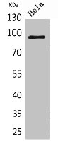
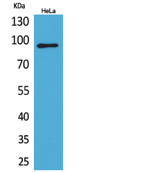
Figure 1. Western blot analysis of CD18/ITGB2 using anti-CD18/ITGB2 antibody (PB9141). Electrophoresis was performed on a 5-20% SDS-PAGE gel at 70V (Stacking gel) / 90V (Resolving gel) for 2-3 hours. The sample well of each lane was loaded with 30 ug of sample under reducing conditions. Lane 1: human Jurkat whole cell lysates, Lane 2: human Hela whole cell lysates. After electrophoresis, proteins were transferred to a nitrocellulose membrane at 150 mA for 50-90 minutes. Blocked the membrane with 5% non-fat milk/TBS for 1.5 hour at RT. The membrane was incubated with rabbit anti-CD18/ITGB2 antigen affinity purified polyclonal antibody (Catalog # PB9141) at 0.5 microg/mL overnight at 4°C, then washed with TBS-0.1%Tween 3 times with 5 minutes each and probed with a goat anti-rabbit IgG-HRP secondary antibody at a dilution of 1:5000 for 1.5 hour at RT. The signal is developed using an Enhanced Chemiluminescent detection (ECL) kit (Catalog # EK1002) with Tanon 5200 system. A specific band was detected for CD18/ITGB2 at approximately 90-100 kDa. The expected band size for CD18/ITGB2 is at 85 kDa.
. CD18/ITGB2 was detected in a paraffin-embedded section of human intestinal cancer tissue. Heat mediated antigen retrieval was performed in EDTA buffer (pH 8.0, epitope retrieval solution). The tissue section was blocked with 10% goat serum. The tissue section was then incubated with 2 microg/ml rabbit anti-CD18/ITGB2 Antibody (PB9141) overnight at 4°C. Peroxidase Conjugated Goat Anti-rabbit IgG was used as secondary antibody and incubated for 30 minutes at 37°C. The tissue section was developed using HRP Conjugated Rabbit IgG Super Vision Assay Kit (Catalog # SV0002) with DAB as the chromogen.")
. CD18/ITGB2 was detected in a paraffin-embedded section of human lung cancer tissue. Heat mediated antigen retrieval was performed in EDTA buffer (pH 8.0, epitope retrieval solution). The tissue section was blocked with 10% goat serum. The tissue section was then incubated with 2 microg/ml rabbit anti-CD18/ITGB2 Antibody (PB9141) overnight at 4°C. Peroxidase Conjugated Goat Anti-rabbit IgG was used as secondary antibody and incubated for 30 minutes at 37°C. The tissue section was developed using HRP Conjugated Rabbit IgG Super Vision Assay Kit (Catalog # SV0002) with DAB as the chromogen.")
. CD18/ITGB2 was detected in a paraffin-embedded section of human tonsil tissue. Heat mediated antigen retrieval was performed in EDTA buffer (pH 8.0, epitope retrieval solution). The tissue section was blocked with 10% goat serum. The tissue section was then incubated with 5 microg/mL rabbit anti-CD18/ITGB2 Antibody (PB9141) overnight at 4°C. DyLight®488 Conjugated Goat Anti-Rabbit IgG (BA1127) was used as secondary antibody at 1:500 dilution and incubated for 30 minutes at 37°C. The section was counterstained with DAPI. Visualize using a fluorescence microscope and filter sets appropriate for the label used.")
Figure 1. Western blot analysis of CD18/ITGB2 using anti-CD18/ITGB2 antibody (PB9141). Electrophoresis was performed on a 5-20% SDS-PAGE gel at 70V (Stacking gel) / 90V (Resolving gel) for 2-3 hours. The sample well of each lane was loaded with 30 ug of sample under reducing conditions. Lane 1: human Jurkat whole cell lysates, Lane 2: human Hela whole cell lysates. After electrophoresis, proteins were transferred to a nitrocellulose membrane at 150 mA for 50-90 minutes. Blocked the membrane with 5% non-fat milk/TBS for 1.5 hour at RT. The membrane was incubated with rabbit anti-CD18/ITGB2 antigen affinity purified polyclonal antibody (Catalog # PB9141) at 0.5 microg/mL overnight at 4°C, then washed with TBS-0.1%Tween 3 times with 5 minutes each and probed with a goat anti-rabbit IgG-HRP secondary antibody at a dilution of 1:5000 for 1.5 hour at RT. The signal is developed using an Enhanced Chemiluminescent detection (ECL) kit (Catalog # EK1002) with Tanon 5200 system. A specific band was detected for CD18/ITGB2 at approximately 90-100 kDa. The expected band size for CD18/ITGB2 is at 85 kDa.
Anti-CD18/ITGB2 Antibody Picoband(r)
PB9141-CARRIER-FREE
ApplicationsImmunoFluorescence, Western Blot, ImmunoHistoChemistry
Product group Antibodies
ReactivityHuman
TargetITGB2
Overview
- SupplierBoster Bio
- Product NameAnti-CD18/ITGB2 Antibody Picoband(r)
- Delivery Days Customer9
- Application Supplier NoteWB: The detection limit for CD18 is approximately 0.25ng/lane under reducing conditions. Tested Species: In-house tested species with positive results. By Heat: Boiling the paraffin sections in 10mM citrate buffer, pH6.0, for 20mins is required for the staining of formalin/paraffin sections. Other applications have not been tested. Optimal dilutions should be determined by end users.
- ApplicationsImmunoFluorescence, Western Blot, ImmunoHistoChemistry
- CertificationResearch Use Only
- ClonalityPolyclonal
- Concentration500 ug/ml
- Gene ID3689
- Target nameITGB2
- Target descriptionintegrin subunit beta 2
- Target synonymsCD18, LAD, LCAMB, LFA-1, MAC-1, MF17, MFI7, integrin beta-2, cell surface adhesion glycoprotein (LFA-1/CR3/P150,959 beta subunit precursor), complement component 3 receptor 3 and 4 subunit, complement receptor C3 beta-subunit, integrin beta chain, beta 2, integrin, beta 2 (complement component 3 receptor 3 and 4 subunit), leukocyte cell adhesion molecule CD18, leukocyte-associated antigens CD18/11A, CD18/11B, CD18/11C
- HostRabbit
- IsotypeIgG
- Protein IDP05107
- Protein NameIntegrin beta-2
- Scientific DescriptionBoster Bio Anti-CD18/ITGB2 Antibody Picoband® catalog # PB9141. Tested in IF, IHC, WB applications. This antibody reacts with Human. The brand Picoband indicates this is a premium antibody that guarantees superior quality, high affinity, and strong signals with minimal background in Western blot applications. Only our best-performing antibodies are designated as Picoband, ensuring unmatched performance.
- ReactivityHuman
- Storage Instruction-20°C,2°C to 8°C
- UNSPSC12352203
Related products
Product group Antibodies
ITGB2 AntibodyCSB-PA005838
ApplicationsWestern Blot, ELISA
ReactivityHuman
TargetITGB2
- SizePrice
Product group Antibodies
ApplicationsImmunoPrecipitation, Western Blot, ImmunoCytoChemistry, ImmunoHistoChemistry
ReactivityMouse, Rat
TargetITGB2
- SizePrice
Product group Antibodies
Anti-ITGB2 AntibodyA101009
ApplicationsWestern Blot, ELISA
ReactivityHuman
- SizePrice
Product group Antibodies
Anti-Human CD18 Antibody, PE136-18066
ApplicationsFlow Cytometry
ReactivityHuman
TargetITGB2
- SizePrice
Product group Antibodies
ApplicationsFlow Cytometry, ImmunoFluorescence
ReactivityHuman
TargetITGB2
- SizePrice
Product group Antibodies

Anti-CD18 [1B4]Ab01201-10.0
ApplicationsFlow Cytometry, ImmunoFluorescence, ImmunoPrecipitation, ELISA, RadioImmunoAssay
ReactivityHuman, Primate
TargetITGB2
- SizePrice
Product group Antibodies

ApplicationsFlow Cytometry
ReactivityMouse
TargetITGB2
- SizePrice

Product group Antibodies
Anti-ITGB2 AntibodyHPA008877
ApplicationsWestern Blot, ImmunoHistoChemistry
ReactivityHuman
TargetITGB2
- SizePrice
Product group Antibodies

ApplicationsWestern Blot, ELISA, ImmunoHistoChemistry
ReactivityHuman
TargetITGB2
- SizePrice