


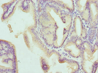
Immunohistochemical staining of human urinary bladder shows strong cytoplasmic positivity in urothelial cells.
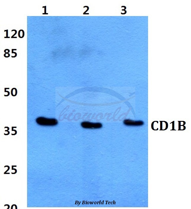
 and CD1B over-expression lysate (Co-expressed with a C-terminal myc-DDK tag (~3.1 kDa) in mammalian HEK293T cells, LY419761).")
Immunohistochemical staining of human urinary bladder shows strong cytoplasmic positivity in urothelial cells.
Anti-CD1B Antibody
HPA021824
ApplicationsWestern Blot, ImmunoHistoChemistry
Product group Antibodies
ReactivityHuman
TargetCD1B
Overview
- SupplierAtlas Antibodies
- Product NameAnti-CD1B Antibody
- Delivery Days Customer4
- ApplicationsWestern Blot, ImmunoHistoChemistry
- CertificationResearch Use Only
- ClonalityPolyclonal
- ConjugateUnconjugated
- Gene ID910
- Target nameCD1B
- Target descriptionCD1b molecule
- Target synonymsCD1, CD1A, R1, T-cell surface glycoprotein CD1b, CD1B antigen, b polypeptide, cortical thymocyte antigen CD1B, differentiation antigen CD1-alpha-3
- HostRabbit
- IsotypeIgG
- Protein IDP29016
- Protein NameT-cell surface glycoprotein CD1b
- Scientific DescriptionRecombinant Protein Epitope Signature Tag (PrEST) antigen sequence
- ReactivityHuman
- Storage Instruction-20°C,2°C to 8°C
- UNSPSC41116161
Datasheet
MSDS
Related products
Product group Antibodies
Anti-CD1B AntibodyA28512
ApplicationsWestern Blot
ReactivityHuman, Mouse, Rat
- SizePrice
Product group Antibodies
Anti-CD1B Antibody144-06551
ApplicationsWestern Blot
ReactivityHuman
TargetCD1B
- SizePrice
Product group Antibodies

CD1B AntibodyLS-C829969
ApplicationsELISA, ImmunoHistoChemistry
ReactivityHuman
TargetCD1B
- SizePrice

Product group Antibodies
Anti-CD1b Antibody Picoband(r)A02158-1-CARRIER-FREE
ApplicationsWestern Blot, ImmunoHistoChemistry
ReactivityHuman
TargetCD1B
- SizePrice
Product group Antibodies
CD1b Monoclonal AntibodyBSM-60395M
ApplicationsFlow Cytometry
ReactivityHuman
TargetCD1B
- SizePrice
Product group Antibodies
CD1B AntibodyCSB-PA004890ESR2HU
ApplicationsELISA, ImmunoHistoChemistry
ReactivityHuman
TargetCD1B
- SizePrice
Product group Antibodies
CD1b antibody [N2C3]GTX112817
ApplicationsWestern Blot
ReactivityHuman
TargetCD1B
- SizePrice
Product group Antibodies
Anti-CD1B AntibodyCAB6551
ApplicationsWestern Blot, ELISA
ReactivityHuman
TargetCD1B
- SizePrice