

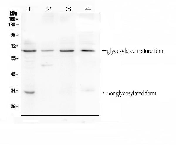
Figure 1. Western blot analysis of CD272 using anti-CD272 antibody (A03149). Electrophoresis was performed on a 5-20% SDS-PAGE gel at 70V (Stacking gel) / 90V (Resolving gel) for 2-3 hours. The sample well of each lane was loaded with 50ug of sample under reducing conditions. Lane 1: human HEK293 whole cell lysate, Lane 2: human Jurkat whole cell lysate, Lane 3: human CCRF-CEM whole cell lysate, Lane 4: mouse thymus tissue lysate. After Electrophoresis, proteins were transferred to a Nitrocellulose membrane at 150mA for 50-90 minutes. Blocked the membrane with 5% Non-fat Milk/ TBS for 1.5 hour at RT. The membrane was incubated with rabbit anti-CD272 antigen affinity purified polyclonal antibody (Catalog # A03149) at 0.5 microg/mL overnight at 4°C, then washed with TBS-0.1%Tween 3 times with 5 minutes each and probed with a goat anti-rabbit IgG-HRP secondary antibody at a dilution of 1:10000 for 1.5 hour at RT. The signal is developed using an Enhanced Chemiluminescent detection (ECL) kit (Catalog # EK1002) with Tanon 5200 system. A specific band was detected for CD272 at approximately 65KD. The expected band size for CD272 is at 33KD.
. CD272/BTLA was detected in paraffin-embedded section of mouse spleen tissues. Heat mediated antigen retrieval was performed in citrate buffer (pH6, epitope retrieval solution) for 20 mins. The tissue section was blocked with 10% goat serum. The tissue section was then incubated with 1microg/ml rabbit anti-CD272/BTLA Antibody (A03149) overnight at 4°C. Biotinylated goat anti-rabbit IgG was used as secondary antibody and incubated for 30 minutes at 37°C. The tissue section was developed using Strepavidin-Biotin-Complex (SABC)(Catalog # SA1022) with DAB as the chromogen.")
. CD272/BTLA was detected in paraffin-embedded section of mouse spleen tissues. Heat mediated antigen retrieval was performed in citrate buffer (pH6, epitope retrieval solution) for 20 mins. The tissue section was blocked with 10% goat serum. The tissue section was then incubated with 1microg/ml rabbit anti-CD272/BTLA Antibody (A03149) overnight at 4°C. Biotinylated goat anti-rabbit IgG was used as secondary antibody and incubated for 30 minutes at 37°C. The tissue section was developed using Strepavidin-Biotin-Complex (SABC)(Catalog # SA1022) with DAB as the chromogen.")
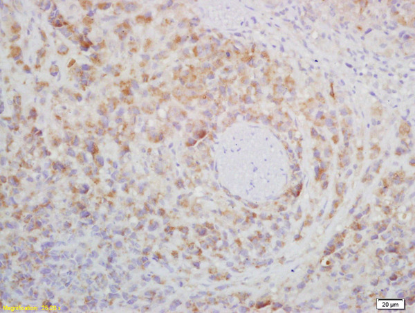
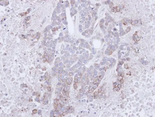
. CD272/BTLA was detected in paraffin-embedded section of human tonsil tissues. Heat mediated antigen retrieval was performed in citrate buffer (pH6, epitope retrieval solution) for 20 mins. The tissue section was blocked with 10% goat serum. The tissue section was then incubated with 1microg/ml rabbit anti-CD272/BTLA Antibody (A03149) overnight at 4°C. Biotinylated goat anti-rabbit IgG was used as secondary antibody and incubated for 30 minutes at 37°C. The tissue section was developed using Strepavidin-Biotin-Complex (SABC)(Catalog # SA1022) with DAB as the chromogen.")
. Overlay histogram showing THP-1 cells stained with A03149 (Blue line). The cells were fixed with 4% paraformaldehyde and blocked with 10% normal goat serum. And then incubated with rabbit anti-CD272/BTLA Antibody (A03149,1microg/1x106 cells) for 30 min at 20°C. DyLight®488 conjugated goat anti-rabbit IgG (BA1127, 5-10microg/1x106 cells) was used as secondary antibody for 30 minutes at 20°C. Isotype control antibody (Green line) was rabbit IgG (1microg/1x106) used under the same conditions. Unlabelled sample (Red line) was also used as a control.")
Figure 1. Western blot analysis of CD272 using anti-CD272 antibody (A03149). Electrophoresis was performed on a 5-20% SDS-PAGE gel at 70V (Stacking gel) / 90V (Resolving gel) for 2-3 hours. The sample well of each lane was loaded with 50ug of sample under reducing conditions. Lane 1: human HEK293 whole cell lysate, Lane 2: human Jurkat whole cell lysate, Lane 3: human CCRF-CEM whole cell lysate, Lane 4: mouse thymus tissue lysate. After Electrophoresis, proteins were transferred to a Nitrocellulose membrane at 150mA for 50-90 minutes. Blocked the membrane with 5% Non-fat Milk/ TBS for 1.5 hour at RT. The membrane was incubated with rabbit anti-CD272 antigen affinity purified polyclonal antibody (Catalog # A03149) at 0.5 microg/mL overnight at 4°C, then washed with TBS-0.1%Tween 3 times with 5 minutes each and probed with a goat anti-rabbit IgG-HRP secondary antibody at a dilution of 1:10000 for 1.5 hour at RT. The signal is developed using an Enhanced Chemiluminescent detection (ECL) kit (Catalog # EK1002) with Tanon 5200 system. A specific band was detected for CD272 at approximately 65KD. The expected band size for CD272 is at 33KD.
Anti-CD272/BTLA Antibody Picoband(r)
A03149-CARRIER-FREE
ApplicationsFlow Cytometry, Western Blot, ImmunoCytoChemistry, ImmunoHistoChemistry, ImmunoHistoChemistry Frozen
Product group Antibodies
ReactivityHuman, Mouse
TargetBTLA
Overview
- SupplierBoster Bio
- Product NameAnti-CD272/BTLA Antibody Picoband(r)
- Delivery Days Customer9
- ApplicationsFlow Cytometry, Western Blot, ImmunoCytoChemistry, ImmunoHistoChemistry, ImmunoHistoChemistry Frozen
- CertificationResearch Use Only
- ClonalityPolyclonal
- Concentration500 ug/ml
- Gene ID151888
- Target nameBTLA
- Target descriptionB and T lymphocyte associated
- Target synonymsBTLA1, CD272, B- and T-lymphocyte attenuator, B- and T-lymphocyte-associated protein
- HostRabbit
- IsotypeIgG
- Protein IDQ7Z6A9
- Protein NameB- and T-lymphocyte attenuator
- Scientific DescriptionBoster Bio Anti-CD272/BTLA Antibody Picoband® catalog # A03149. Tested in Flow Cytometry, IHC, IHC-F, ICC, WB applications. This antibody reacts with Human, Mouse. The brand Picoband indicates this is a premium antibody that guarantees superior quality, high affinity, and strong signals with minimal background in Western blot applications. Only our best-performing antibodies are designated as Picoband, ensuring unmatched performance.
- ReactivityHuman, Mouse
- Storage Instruction-20°C,2°C to 8°C
- UNSPSC12352203
Related products
Product group Antibodies
anti-BTLA (human), mAb (6F4)AG-20B-0049
ApplicationsFlow Cytometry, ELISA
ReactivityHuman
TargetBTLA
- SizePrice
Product group Antibodies
Anti-BTLA Antibody144-08377
ApplicationsWestern Blot
ReactivityHuman, Mouse, Rat
TargetBTLA
- SizePrice
Product group Antibodies

BTLA / CD272 Antibody (clone PK18.6)LS-C770041
ApplicationsFlow Cytometry
ReactivityMouse
TargetBTLA
- SizePrice
Product group Antibodies
CD272 Polyclonal AntibodyBS-0624R
ApplicationsFlow Cytometry, ImmunoFluorescence, Western Blot, ELISA, ImmunoCytoChemistry, ImmunoHistoChemistry, ImmunoHistoChemistry Frozen, ImmunoHistoChemistry Paraffin
ReactivityHuman, Mouse, Rat
TargetBTLA
- SizePrice
Product group Antibodies
BTLA AntibodyCSB-PA007471
ApplicationsWestern Blot, ELISA
ReactivityHuman
TargetBTLA
- SizePrice
Product group Antibodies

Goat anti-BTLAEB09035
ApplicationsWestern Blot, ELISA
ReactivityHuman, Mouse, Rat
TargetBTLA
- SizePrice
Product group Antibodies
ApplicationsImmunoPrecipitation, Western Blot, ImmunoCytoChemistry, ImmunoHistoChemistry
TargetBTLA
- SizePrice

Product group Antibodies
Anti-BTLA AntibodyHPA062029
ApplicationsImmunoHistoChemistry
ReactivityHuman
TargetBTLA
- SizePrice
Product group Antibodies
CD272 antibodyGTX112783
ApplicationsWestern Blot, ImmunoHistoChemistry, ImmunoHistoChemistry Paraffin
ReactivityHuman
TargetBTLA
- SizePrice