


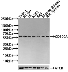
Figure 1. Western blot analysis of CD300A using anti-CD300A antibody (A05953-1). Electrophoresis was performed on a 5-20% SDS-PAGE gel at 70V (Stacking gel) / 90V (Resolving gel) for 2-3 hours. The sample well of each lane was loaded with 30 ug of sample under reducing conditions. Lane 1: human HL-60 whole cell lysates, Lane 2: human Raji whole cell lysates, Lane 3: rat PC-12 whole cell lysates, Lane 4: rat C6 whole cell lysates, Lane 5: mouse Raw264.7 whole cell lysates, Lane 6: mouse J774A.1 whole cell lysates. After electrophoresis, proteins were transferred to a nitrocellulose membrane at 150 mA for 50-90 minutes. Blocked the membrane with 5% non-fat milk/TBS for 1.5 hour at RT. The membrane was incubated with rabbit anti-CD300A antigen affinity purified polyclonal antibody (Catalog # A05953-1) at 0.5 microg/mL overnight at 4°C, then washed with TBS-0.1%Tween 3 times with 5 minutes each and probed with a goat anti-rabbit IgG-HRP secondary antibody at a dilution of 1:5000 for 1.5 hour at RT. The signal is developed using an Enhanced Chemiluminescent detection (ECL) kit (Catalog # EK1002) with Tanon 5200 system. A specific band was detected for CD300A at approximately 68 kDa. The expected band size for CD300A is at 33 kDa.
. Overlay histogram showing U20S cells stained with A05953-1 (Blue line). The cells were fixed with 4% paraformaldehyde and blocked with 10% normal goat serum. And then incubated with rabbit anti-CD300A Antibody (A05953-1, 1 microg/1x106 cells) for 30 min at 20°C. DyLight®488 conjugated goat anti-rabbit IgG (BA1127, 5-10 microg/1x106 cells) was used as secondary antibody for 30 minutes at 20°C. Isotype control antibody (Green line) was rabbit IgG (1 microg/1x106) used under the same conditions. Unlabelled sample (Red line) was also used as a control.")
Figure 1. Western blot analysis of CD300A using anti-CD300A antibody (A05953-1). Electrophoresis was performed on a 5-20% SDS-PAGE gel at 70V (Stacking gel) / 90V (Resolving gel) for 2-3 hours. The sample well of each lane was loaded with 30 ug of sample under reducing conditions. Lane 1: human HL-60 whole cell lysates, Lane 2: human Raji whole cell lysates, Lane 3: rat PC-12 whole cell lysates, Lane 4: rat C6 whole cell lysates, Lane 5: mouse Raw264.7 whole cell lysates, Lane 6: mouse J774A.1 whole cell lysates. After electrophoresis, proteins were transferred to a nitrocellulose membrane at 150 mA for 50-90 minutes. Blocked the membrane with 5% non-fat milk/TBS for 1.5 hour at RT. The membrane was incubated with rabbit anti-CD300A antigen affinity purified polyclonal antibody (Catalog # A05953-1) at 0.5 microg/mL overnight at 4°C, then washed with TBS-0.1%Tween 3 times with 5 minutes each and probed with a goat anti-rabbit IgG-HRP secondary antibody at a dilution of 1:5000 for 1.5 hour at RT. The signal is developed using an Enhanced Chemiluminescent detection (ECL) kit (Catalog # EK1002) with Tanon 5200 system. A specific band was detected for CD300A at approximately 68 kDa. The expected band size for CD300A is at 33 kDa.
Anti-CD300A Antibody Picoband(r)
A05953-1-CARRIER-FREE
ApplicationsFlow Cytometry, Western Blot, ELISA
Product group Antibodies
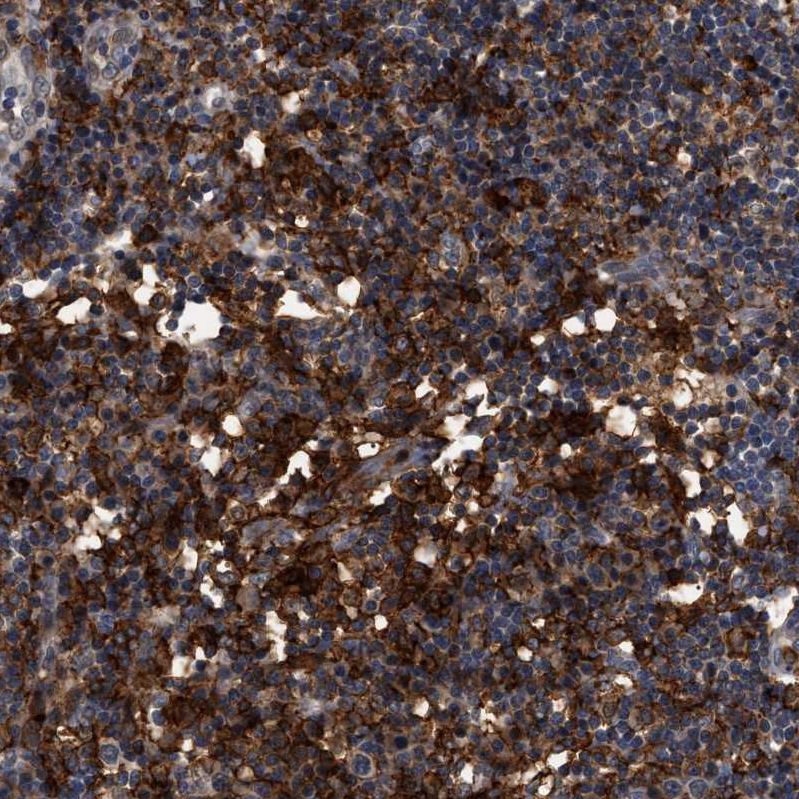
ReactivityHuman, Mouse, Rat
TargetCD300A
Overview
- SupplierBoster Bio
- Product NameAnti-CD300A Antibody Picoband(r)
- Delivery Days Customer9
- ApplicationsFlow Cytometry, Western Blot, ELISA
- CertificationResearch Use Only
- ClonalityPolyclonal
- Concentration500 ug/ml
- Gene ID11314
- Target nameCD300A
- Target descriptionCD300a molecule
- Target synonymsCLM-8, CMRF-35-H9, CMRF-35H, CMRF35-H, CMRF35-H9, CMRF35H, CMRF35H9, IGSF12, IRC1, IRC1/IRC2, IRC2, IRp60, CMRF35-like molecule 8, CD300 antigen-like family member A, CD300a antigen, CMRF35H leukocyte immunoglobulin-like receptor, NK inhibitory receptor, immunoglobulin superfamily member 12, inhibitory receptor protein 60, leukocyte membrane antigen
- HostRabbit
- IsotypeIgG
- Protein IDQ9UGN4
- Protein NameCMRF35-like molecule 8
- Scientific DescriptionBoster Bio Anti-CD300A Antibody Picoband® catalog # A10247-2. Tested in WB, Flow Cytometry, ELISA applications. This antibody reacts with Human, Mouse, Rat. The brand Picoband indicates this is a premium antibody that guarantees superior quality, high affinity, and strong signals with minimal background in Western blot applications. Only our best-performing antibodies are designated as Picoband, ensuring unmatched performance.
- ReactivityHuman, Mouse, Rat
- Storage Instruction-20°C,2°C to 8°C
- UNSPSC12352203
Related products
Product group Antibodies
IGSF12 Polyclonal AntibodyBS-9202R
ApplicationsImmunoFluorescence, Western Blot, ImmunoHistoChemistry, ImmunoHistoChemistry Paraffin
ReactivityHuman
TargetCD300A
- SizePrice
Product group Antibodies
CD300A AntibodyCSB-PA887023ESR1HU
ApplicationsWestern Blot, ELISA, ImmunoHistoChemistry
ReactivityHuman, Mouse
TargetCD300A
- SizePrice
Product group Antibodies
Cd300A Polyclonal AntibodyCAC10862
ApplicationsWestern Blot, ELISA, ImmunoHistoChemistry
ReactivityMouse
TargetCD300A
- SizePrice
Product group Antibodies

CD300A AntibodyLS-C496719
ApplicationsWestern Blot
ReactivityHuman
TargetCD300A
- SizePrice
Product group Antibodies
CD300a antibody [MEM-260] (PE)GTX79961
ApplicationsFlow Cytometry
ReactivityHuman
TargetCD300A
- SizePrice
Product group Antibodies
Anti-CD300A AntibodyHPA011645
ApplicationsImmunoHistoChemistry
ReactivityHuman
TargetCD300A
- SizePrice
Product group Antibodies
Anti-CD300A AntibodyCAB10006
ApplicationsWestern Blot, ELISA
ReactivityHuman
TargetCD300A
- SizePrice