

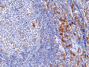
Immunohistochemical staining of formalin fixed and paraffin embedded human tonsil tissue section using anti-CD33 rabbit monoclonal antibody (Clone RM398) at a 1:100 dilution.
Immunohistochemical staining of formalin fixed and paraffin embedded human tonsil tissue section using anti-CD33 rabbit monoclonal antibody (Clone RM398) at a 1:100 dilution.
anti-CD33 (human), Rabbit Monoclonal (RM398)
REV-31-1284-00
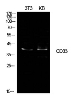
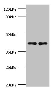
ApplicationsWestern Blot, ImmunoHistoChemistry
Product group Antibodies
ReactivityHuman
TargetCD33
Overview
- SupplierRevMAb Biosciences
- Product Nameanti-CD33 (human), Rabbit Monoclonal (RM398)
- Delivery Days Customer10
- ApplicationsWestern Blot, ImmunoHistoChemistry
- CertificationResearch Use Only
- ClonalityMonoclonal
- Clone IDRM398
- Gene ID945
- Target nameCD33
- Target descriptionCD33 molecule
- Target synonymsCD33rSiglec, SIGLEC-3, SIGLEC3, p67, myeloid cell surface antigen CD33, CD33 antigen (gp67), CD33 molecule transcript, gp67, sialic acid-binding Ig-like lectin 3
- HostRabbit
- IsotypeIgG
- Protein IDP20138
- Protein NameMyeloid cell surface antigen CD33
- Scientific DescriptionCD33 is a transmembrane protein of the sialic acid-binding immunoglobulin-like lectin (Siglec) family. It belongs to the immunoreceptor tyrosine-based inhibitory motif (ITIM)-containing molecules able of recruiting protein tyrosine phosphatases SHP-1 and SHP-2 to signal assemblies, and these ITIMs are used for ubiquitin-mediated removal of the receptor from the cell surface. CD33 is expressed on cells of myelomonocytic lineage, binds sialic acid residues in N- and O-glycans on cell surfaces, and is a therapeutic target for acute myeloid leukemia. CD33 is found on granulocyte and macrophage precursors in the bone marrow, but is not on pluripotent stem cells. CD33 is also a useful marker for peripheral monocytes. CD33 is useful for distinguishing myelogenous leukemia cells from lymphoid or erythroid leukemias. Diseases associated with CD33 dysfunction include gallbladder lymphoma and extracutaneous mastocytoma. - Recombinant Antibody. This antibody reacts to human CD33. Applications: WB, IHC. Source: Rabbit. Liquid. 50% Glycerol/PBS with 1% BSA and 0.09% sodium azide. CD33 is a transmembrane protein of the sialic acid-binding immunoglobulin-like lectin (Siglec) family. It belongs to the immunoreceptor tyrosine-based inhibitory motif (ITIM)-containing molecules able of recruiting protein tyrosine phosphatases SHP-1 and SHP-2 to signal assemblies, and these ITIMs are used for ubiquitin-mediated removal of the receptor from the cell surface. CD33 is expressed on cells of myelomonocytic lineage, binds sialic acid residues in N- and O-glycans on cell surfaces, and is a therapeutic target for acute myeloid leukemia. CD33 is found on granulocyte and macrophage precursors in the bone marrow, but is not on pluripotent stem cells. CD33 is also a useful marker for peripheral monocytes. CD33 is useful for distinguishing myelogenous leukemia cells from lymphoid or erythroid leukemias. Diseases associated with CD33 dysfunction include gallbladder lymphoma and extracutaneous mastocytoma.
- ReactivityHuman
- Storage Instruction-20°C,2°C to 8°C
- UNSPSC41116161
Datasheet
Related products
Product group Antibodies

Anti-CD33 [hP67.6 (Gemtuzumab)]Ab00283-1.1
ApplicationsFlow Cytometry, ImmunoFluorescence
ReactivityHuman
TargetCD33
- SizePrice
Product group Antibodies
Anti-CD33 AntibodyA101517
ApplicationsWestern Blot, ELISA
ReactivityHuman
- SizePrice
Product group Antibodies
CD33 Polyclonal AntibodyBS-2042R
ApplicationsFlow Cytometry, ImmunoFluorescence, Western Blot, ELISA, ImmunoCytoChemistry, ImmunoHistoChemistry, ImmunoHistoChemistry Frozen, ImmunoHistoChemistry Paraffin
ReactivityHuman
TargetCD33
- SizePrice
Product group Antibodies
Cd33 Polyclonal AntibodyCAC11135
ApplicationsWestern Blot, ELISA, ImmunoHistoChemistry
TargetCD33
- SizePrice
Product group Antibodies
CD33 AntibodyCSB-PA004925ESR1HU
ApplicationsWestern Blot, ELISA, ImmunoHistoChemistry
ReactivityHuman
TargetCD33
- SizePrice
Product group Antibodies
CD33 antibody [WM53] (PE-Cy7)GTX00477-10
ApplicationsFlow Cytometry
ReactivityHuman, Primate
TargetCD33
- SizePrice
Product group Antibodies

MERS-CoV Nucleoprotein AntibodyLS-C488273
ApplicationsELISA
ReactivityVirus
- SizePrice

Product group Antibodies
Anti-CD33 AntibodyHPA035832
ApplicationsImmunoHistoChemistry
ReactivityHuman
TargetCD33
- SizePrice