


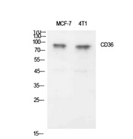
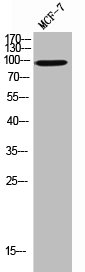
Figure 1. Western blot analysis of CD36 using anti-CD36 antibody (A01189-1). Electrophoresis was performed on a 5-20% SDS-PAGE gel at 70V (Stacking gel) / 90V (Resolving gel) for 2-3 hours. The sample well of each lane was loaded with 30 ug of sample under reducing conditions. Lane 1: human HL-60 whole cell lysates, Lane 2: human HepG2 whole cell lysates. After electrophoresis, proteins were transferred to a nitrocellulose membrane at 150 mA for 50-90 minutes. Blocked the membrane with 5% non-fat milk/TBS for 1.5 hour at RT. The membrane was incubated with rabbit anti-CD36 antigen affinity purified polyclonal antibody (Catalog # A01189-1) at 0.5 microg/mL overnight at 4°C, then washed with TBS-0.1%Tween 3 times with 5 minutes each and probed with a goat anti-rabbit IgG-HRP secondary antibody at a dilution of 1:5000 for 1.5 hour at RT. The signal is developed using an Enhanced Chemiluminescent detection (ECL) kit (Catalog # EK1002) with Tanon 5200 system. A specific band was detected for CD36 at approximately 88 kDa. The expected band size for CD36 is at 53 kDa.
. Overlay histogram showing HEL cells stained with A01189-1 (Blue line). The cells were blocked with 10% normal goat serum. And then incubated with rabbit anti-CD36 Antibody (A01189-1, 1 microg/1x106 cells) for 30 min at 20°C. DyLight®488 conjugated goat anti-rabbit IgG (BA1127, 5-10 microg/1x106 cells) was used as secondary antibody for 30 minutes at 20°C. Isotype control antibody (Green line) was rabbit IgG (1 microg/1x106) used under the same conditions. Unlabelled sample (Red line) was also used as a control.")
Figure 1. Western blot analysis of CD36 using anti-CD36 antibody (A01189-1). Electrophoresis was performed on a 5-20% SDS-PAGE gel at 70V (Stacking gel) / 90V (Resolving gel) for 2-3 hours. The sample well of each lane was loaded with 30 ug of sample under reducing conditions. Lane 1: human HL-60 whole cell lysates, Lane 2: human HepG2 whole cell lysates. After electrophoresis, proteins were transferred to a nitrocellulose membrane at 150 mA for 50-90 minutes. Blocked the membrane with 5% non-fat milk/TBS for 1.5 hour at RT. The membrane was incubated with rabbit anti-CD36 antigen affinity purified polyclonal antibody (Catalog # A01189-1) at 0.5 microg/mL overnight at 4°C, then washed with TBS-0.1%Tween 3 times with 5 minutes each and probed with a goat anti-rabbit IgG-HRP secondary antibody at a dilution of 1:5000 for 1.5 hour at RT. The signal is developed using an Enhanced Chemiluminescent detection (ECL) kit (Catalog # EK1002) with Tanon 5200 system. A specific band was detected for CD36 at approximately 88 kDa. The expected band size for CD36 is at 53 kDa.
Anti-CD36 Antibody Picoband(r)
A01189-1-CARRIER-FREE
ApplicationsFlow Cytometry, Western Blot
Product group Antibodies
ReactivityHuman, Mouse, Rat
TargetCD36
Overview
- SupplierBoster Bio
- Product NameAnti-CD36 Antibody Picoband(r)
- Delivery Days Customer9
- ApplicationsFlow Cytometry, Western Blot
- CertificationResearch Use Only
- ClonalityPolyclonal
- Concentration500 ug/ml
- Gene ID948
- Target nameCD36
- Target descriptionCD36 molecule (CD36 blood group)
- Target synonymsBDPLT10, CHDS7, FAT, GP3B, GP4, GPIV, PASIV, SCARB3, platelet glycoprotein 4, CD36 antigen (collagen type I receptor, thrombospondin receptor), CD36 molecule (CD36 blood group) transcript, CD36 molecule (thrombospondin receptor), GPIIIB, PAS IV, PAS-4 protein, cluster determinant 36, fatty acid translocase, glycoprotein IIIb, leukocyte differentiation antigen CD36, platelet glycoprotein IV, scavenger receptor class B, member 3
- HostRabbit
- IsotypeIgG
- Protein IDP16671
- Protein NamePlatelet glycoprotein 4
- Scientific DescriptionBoster Bio Anti-CD36 Antibody Picoband® catalog # A01189-1. Tested in ELISA, Flow Cytometry, WB applications. This antibody reacts with Human, Mouse, Rat. The brand Picoband indicates this is a premium antibody that guarantees superior quality, high affinity, and strong signals with minimal background in Western blot applications. Only our best-performing antibodies are designated as Picoband, ensuring unmatched performance.
- ReactivityHuman, Mouse, Rat
- Storage Instruction-20°C,2°C to 8°C
- UNSPSC12352203
References
- Kuda O, Pietka TA, Demianova Z, et al. Sulfo-N-succinimidyl oleate (SSO) inhibits fatty acid uptake and signaling for intracellular calcium via binding CD36 lysine 164: SSO also inhibits oxidized low density lipoprotein uptake by macrophages. J Biol Chem. 2013,288(22):15547-55. doi: 10.1074/jbc.M113.473298Read this paper
- Tandon NN, Kralisz U, Jamieson GA. Identification of glycoprotein IV (CD36) as a primary receptor for platelet-collagen adhesion. J Biol Chem. 1989,264(13):7576-83.Read this paper
- Silverstein RL, Baird M, Lo SK, et al. Sense and antisense cDNA transfection of CD36 (glycoprotein IV) in melanoma cells. Role of CD36 as a thrombospondin receptor. J Biol Chem. 1992,267(23):16607-12.Read this paper
Related products
Product group Antibodies
Anti-CD36 AntibodyA98246
ApplicationsWestern Blot, ELISA
ReactivityHuman, Mouse, Rat
- SizePrice
Product group Antibodies

Anti-CD36 [185.IG2], Human IgG1, kappaAB04254-10.0
ApplicationsImmunoFluorescence, ImmunoPrecipitation, ImmunoHistoChemistry, Neutralisation/Blocking
ReactivityHuman, Mouse
TargetCD36
- SizePrice
Product group Antibodies

CD36 Antibody (clone 5-271, FITC)LS-C811575
ApplicationsFlow Cytometry
ReactivityHuman
TargetCD36
- SizePrice
Product group Antibodies
CD36 Recombinant Antibody, AbBy Fluor-350 ConjugatedBSM-61310R-BF350
ApplicationsFlow Cytometry, ImmunoFluorescence, Western Blot
TargetCD36
- SizePrice
Product group Antibodies
CD36 AntibodyCSB-PA006183
ApplicationsWestern Blot, ELISA
ReactivityHuman, Mouse, Rat
TargetCD36
- SizePrice
Product group Antibodies
Cd36 Polyclonal AntibodyCAC09109
ApplicationsImmunoFluorescence, Western Blot, ELISA, ImmunoHistoChemistry
ReactivityMouse
TargetCD36
- SizePrice
![Untreated (–) and treated (+) THP-1 whole cell extracts (30 μg) were separated by 7.5% SDS-PAGE, and the membrane was blotted with CD36 antibody [C1C3] (GTX100642) diluted at 1:1000. The HRP-conjugated anti-rabbit IgG antibody (GTX213110-01) was used to detect the primary antibody, and the signal was developed with Trident ECL plus-Enhanced.](https://www.genetex.com/upload/website/prouct_img/normal/GTX100642/GTX100642_42298_20220923_WB_treatment_PMA_22092622_830.webp)
Product group Antibodies
CD36 antibody [C1C3]GTX100642
ApplicationsImmunoFluorescence, Western Blot, ImmunoCytoChemistry, ImmunoHistoChemistry, ImmunoHistoChemistry Paraffin
ReactivityHuman, Mouse
TargetCD36
- SizePrice

Product group Antibodies
Anti-CD36 AntibodyHPA002018
ApplicationsImmunoCytoChemistry, ImmunoHistoChemistry
ReactivityHuman
TargetCD36
- SizePrice