


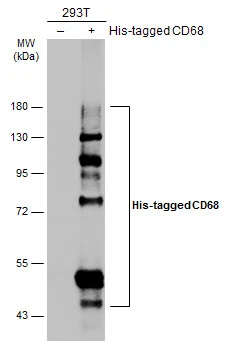
Figure 1. Western blot analysis of CD68 using anti-CD68 antibody (PA1518). Electrophoresis was performed on a 5-20% SDS-PAGE gel at 70V (Stacking gel) / 90V (Resolving gel) for 2-3 hours. The sample well of each lane was loaded with 30 ug of sample under reducing conditions. Lane 1: rat spleen tissue lysates, Lane 2: mouse spleen tissue lysates, Lane 3: mouse RAW264.7 whole cell lysates. After electrophoresis, proteins were transferred to a nitrocellulose membrane at 150 mA for 50-90 minutes. Blocked the membrane with 5% non-fat milk/TBS for 1.5 hour at RT. The membrane was incubated with rabbit anti-CD68 antigen affinity purified polyclonal antibody (Catalog # PA1518) at 0.5 microg/mL overnight at 4°C, then washed with TBS-0.1%Tween 3 times with 5 minutes each and probed with a goat anti-rabbit IgG-HRP secondary antibody at a dilution of 1:5000 for 1.5 hour at RT. The signal is developed using an Enhanced Chemiluminescent detection (ECL) kit (Catalog # EK1002) with Tanon 5200 system. A specific band was detected for CD68 at approximately 90-100 kDa. The expected band size for CD68 is at 37 kDa.
. CD68 was detected in a paraffin-embedded section of mouse liver tissue. Heat mediated antigen retrieval was performed in EDTA buffer (pH 8.0, epitope retrieval solution). The tissue section was blocked with 10% goat serum. The tissue section was then incubated with 2 microg/ml rabbit anti-CD68 Antibody (PA1518) overnight at 4°C. Peroxidase Conjugated Goat Anti-rabbit IgG was used as secondary antibody and incubated for 30 minutes at 37°C. The tissue section was developed using HRP Conjugated Rabbit IgG Super Vision Assay Kit (Catalog # SV0002) with DAB as the chromogen.")
. CD68 was detected in a paraffin-embedded section of mouse spleen tissue. Heat mediated antigen retrieval was performed in EDTA buffer (pH 8.0, epitope retrieval solution). The tissue section was blocked with 10% goat serum. The tissue section was then incubated with 2 microg/ml rabbit anti-CD68 Antibody (PA1518) overnight at 4°C. Peroxidase Conjugated Goat Anti-rabbit IgG was used as secondary antibody and incubated for 30 minutes at 37°C. The tissue section was developed using HRP Conjugated Rabbit IgG Super Vision Assay Kit (Catalog # SV0002) with DAB as the chromogen.")
. CD68 was detected in a paraffin-embedded section of rat spleen tissue. Heat mediated antigen retrieval was performed in EDTA buffer (pH 8.0, epitope retrieval solution). The tissue section was blocked with 10% goat serum. The tissue section was then incubated with 2 microg/ml rabbit anti-CD68 Antibody (PA1518) overnight at 4°C. Peroxidase Conjugated Goat Anti-rabbit IgG was used as secondary antibody and incubated for 30 minutes at 37°C. The tissue section was developed using HRP Conjugated Rabbit IgG Super Vision Assay Kit (Catalog # SV0002) with DAB as the chromogen.")
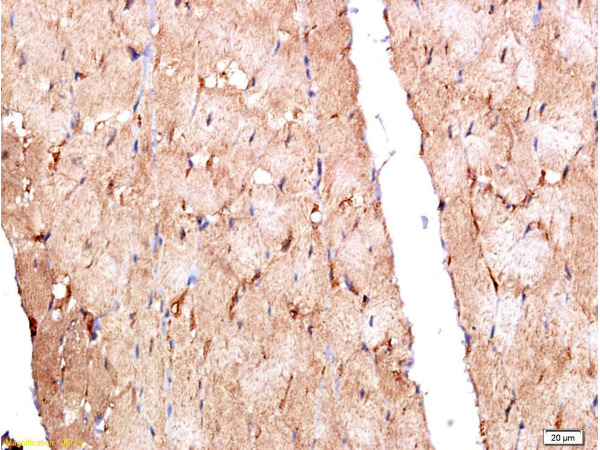
. CD68 was detected in a paraffin-embedded section of rat lung tissue. Heat mediated antigen retrieval was performed in EDTA buffer (pH 8.0, epitope retrieval solution). The tissue section was blocked with 10% goat serum. The tissue section was then incubated with 2 microg/ml rabbit anti-CD68 Antibody (PA1518) overnight at 4°C. Peroxidase Conjugated Goat Anti-rabbit IgG was used as secondary antibody and incubated for 30 minutes at 37°C. The tissue section was developed using HRP Conjugated Rabbit IgG Super Vision Assay Kit (Catalog # SV0002) with DAB as the chromogen.")
. CD68 was detected in a paraffin-embedded section of rat lung tissue. Heat mediated antigen retrieval was performed in EDTA buffer (pH 8.0, epitope retrieval solution). The tissue section was blocked with 10% goat serum. The tissue section was then incubated with 2 microg/ml rabbit anti-CD68 Antibody (PA1518) overnight at 4°C. Peroxidase Conjugated Goat Anti-rabbit IgG was used as secondary antibody and incubated for 30 minutes at 37°C. The tissue section was developed using HRP Conjugated Rabbit IgG Super Vision Assay Kit (Catalog # SV0002) with DAB as the chromogen.")
. CD68 was detected in a paraffin-embedded section of rat liver tissue. Heat mediated antigen retrieval was performed in EDTA buffer (pH 8.0, epitope retrieval solution). The tissue section was blocked with 10% goat serum. The tissue section was then incubated with 2 microg/ml rabbit anti-CD68 Antibody (PA1518) overnight at 4°C. Peroxidase Conjugated Goat Anti-rabbit IgG was used as secondary antibody and incubated for 30 minutes at 37°C. The tissue section was developed using HRP Conjugated Rabbit IgG Super Vision Assay Kit (Catalog # SV0002) with DAB as the chromogen.")
. CD68 was detected in a paraffin-embedded section of mouse spleen tissue. Heat mediated antigen retrieval was performed in EDTA buffer (pH 8.0, epitope retrieval solution). The tissue section was blocked with 10% goat serum. The tissue section was then incubated with 5 microg/mL rabbit anti-CD68 Antibody (PA1518) overnight at 4°C. DyLight®488 Conjugated Goat Anti-Rabbit IgG (BA1127)) was used as secondary antibody at 1:500 dilution and incubated for 30 minutes at 37°C. The section was counterstained with DAPI. Visualize using a fluorescence microscope and filter sets appropriate for the label used.")
. Overlay histogram showing RAW264.7 cells stained with PA1518 (Blue line). The cells were fixed with 4% paraformaldehyde and blocked with 10% normal goat serum. And then incubated with rabbit anti-CD68 Antibody (PA1518, 1 microg/1x106 cells) for 30 min at 20°C. DyLight®488 conjugated goat anti-rabbit IgG (BA1127, 5-10 microg/1x106 cells) was used as secondary antibody for 30 minutes at 20°C. Isotype control antibody (Green line) was rabbit IgG (1 microg/1x106) used under the same conditions. Unlabelled sample without incubation with primary antibody and secondary antibody (Red line) was used as a blank control.")
Figure 1. Western blot analysis of CD68 using anti-CD68 antibody (PA1518). Electrophoresis was performed on a 5-20% SDS-PAGE gel at 70V (Stacking gel) / 90V (Resolving gel) for 2-3 hours. The sample well of each lane was loaded with 30 ug of sample under reducing conditions. Lane 1: rat spleen tissue lysates, Lane 2: mouse spleen tissue lysates, Lane 3: mouse RAW264.7 whole cell lysates. After electrophoresis, proteins were transferred to a nitrocellulose membrane at 150 mA for 50-90 minutes. Blocked the membrane with 5% non-fat milk/TBS for 1.5 hour at RT. The membrane was incubated with rabbit anti-CD68 antigen affinity purified polyclonal antibody (Catalog # PA1518) at 0.5 microg/mL overnight at 4°C, then washed with TBS-0.1%Tween 3 times with 5 minutes each and probed with a goat anti-rabbit IgG-HRP secondary antibody at a dilution of 1:5000 for 1.5 hour at RT. The signal is developed using an Enhanced Chemiluminescent detection (ECL) kit (Catalog # EK1002) with Tanon 5200 system. A specific band was detected for CD68 at approximately 90-100 kDa. The expected band size for CD68 is at 37 kDa.
Anti-Macrosialin CD68 Antibody Picoband(r)
PA1518
ApplicationsFlow Cytometry, ImmunoFluorescence, Western Blot, ImmunoHistoChemistry
Product group Antibodies
ReactivityHamster, Mouse, Rat
TargetCd68
Overview
- SupplierBoster Bio
- Product NameAnti-Macrosialin CD68 Antibody Picoband(r)
- Delivery Days Customer9
- Application Supplier NoteTested Species: In-house tested species with positive results. Predicted Species: Species predicted to be fit for the product based on sequence similarities. By Heat: Boiling the paraffin sections in 10mM citrate buffer, pH6.0, for 20mins is required for the staining of formalin/paraffin sections. Other applications have not been tested. Optimal dilutions should be determined by end users.
- ApplicationsFlow Cytometry, ImmunoFluorescence, Western Blot, ImmunoHistoChemistry
- Applications SupplierIHP, IHF, WB, IHC
- CertificationResearch Use Only
- ClonalityPolyclonal
- Concentration500 ug/ml
- Gene ID12514
- Target nameCd68
- Target descriptionCD68 antigen
- Target synonymsLamp4, Scard1, gp110, macrosialin
- HostRabbit
- IsotypeIgG
- Protein IDP31996
- Protein NameMacrosialin
- Scientific DescriptionSuperstar antibody: Bosters anti mouse CD68 antibody (PA1518) is among its top 10 bestselling antibodies, and one of the most cited mouse/rat CD68 antibodies on the market. Because of this antibodys high demand, we have subsequently developed a rabbit monoclonal anti CD68 antibody (M00602-1) targetting a similar epitope. High specificity: PA1518 reacts with murine macrosialin/CD68, and has been validated with Western blotting and confirmed its stellar specificity. CD68 is a highly glycosylated protein, and its expected western blot molecular weight is between 80kDa to 110kDa, depending on glycosylation level. PA1518s observed MW in WB is near 90-100kDa. Testing on negative control tissues showed no significant bands (images available on request) or staining. See more info in the positive and negative control design section. Based on immunogen sequence homology, this antibody is not expected to cross react with other proteins from the LAMP family, which complies with QC testing observations. Great for CD68 IHC: This antibody produces clean and specific immunobiological stains in both mouse and rat tissues. Our QC team has validated it on spleen and liver tissues of both mice and rats. Click product images for more details on experiment conditions. More about CD68: CD68 is a cell surface marker often used to identify macrophages and other cell types in the monocyte lineage. It is a transmembrane protein that binds to electins and selectins and plays a role in macrophage homing movement. Check out the CD68 biomarker page for more information on CD68 and view all CD68 antibodies, ELISA kits and proteins. The brand Picoband indicates this is a premium antibody that guarantees superior quality, high affinity, and strong signals with minimal background in Western blot applications. Only our best-performing antibodies are designated as Picoband, ensuring unmatched performance.
- ReactivityHamster, Mouse, Rat
- Reactivity SupplierMouse, Rat, Hamster
- Storage Instruction-20°C,2°C to 8°C
- UNSPSC12352203
References
- Lei J, Shu Z, Zhu H, et al. AMPK Regulates M1 Macrophage Polarization through the JAK2/STAT3 Signaling Pathway to Attenuate Airway Inflammation in Obesity-Related Asthma. Inflammation. 2025,48(1):372-392. doi: 10.1007/s10753-024-02070-xRead this paper
- Zhang MX, Hong H, Shi Y, et al. A Pilot Study on a Possible Mechanism behind Olfactory Dysfunction in Parkinson's Disease: The Association of TAAR1 Downregulation with Neuronal Loss and Inflammation along Olfactory Pathway. Brain Sci. 2024,14(4). doi: 10.3390/brainsci14040300Read this paper
- Cao C, Wu R, Wang S, et al. Elucidating the changes in the heterogeneity and function of radiation-induced cardiac macrophages using single-cell RNA sequencing. Front Immunol. 2024,15:1363278. doi: 10.3389/fimmu.2024.1363278Read this paper
- Yan Z, Shi Y, Yang R, et al. ELABELA-derived peptide ELA13 attenuates kidney fibrosis by inhibiting the Smad and ERK signaling pathways. J Zhejiang Univ Sci B. 2024,25(4):341-353. doi: 10.1631/jzus.B2300033Read this paper
- Yu C, Zhu Q, Ma C, et al. Major vault protein regulates tumor-associated macrophage polarization through interaction with signal transducer and activator of transcription 6. Front Immunol. 2023,14:1289795. doi: 10.3389/fimmu.2023.1289795Read this paper
- Yu W, Ding J, Chen J, et al. Magnesium Ion-Doped Mesoporous Bioactive Glasses Loaded with Gallic Acid Against Myocardial Ischemia/Reperfusion Injury by Affecting the Biological Functions of Multiple Cells. Int J Nanomedicine. 2024,19:347-366. doi: 10.2147/IJN.S444751Read this paper
- Lv J, Wang J, Zeng Y, et al. In vitro chemical treatment of silk increases the expression of pro-inflammatory factors and facilitates degradation in rats. J Appl Biomater Funct Mater. 2024,22:22808000231222704. doi: 10.1177/22808000231222704Read this paper
- Wang J, Wu W, Yuan T, et al. Tumor-associated macrophages and PD-L1 in prostate cancer: a possible key to unlocking immunotherapy efficacy. Aging (Albany NY). 2024,16(1):445-465. doi: 10.18632/aging.205378Read this paper
- Kuang Q, Gao L, Feng L, et al. Toxicological effects of microplastics in renal ischemia-reperfusion injury. Environ Toxicol. 2024,39(4):2350-2362. doi: 10.1002/tox.24115Read this paper
- Lan T, Chen B, Hu X, et al. Tianhuang formula ameliorates liver fibrosis by inhibiting CCL2-CCR2 axis and MAPK/NF-κB signaling pathway. J Ethnopharmacol. 2024,321:117516. doi: 10.1016/j.jep.2023.117516Read this paper
Datasheet
MSDS
Related products
Product group Antibodies

Anti-CD68 [FA-11]Ab01113-2.0
ApplicationsFlow Cytometry, ImmunoPrecipitation, ImmunoHistoChemistry
ReactivityMouse
TargetCd68
- SizePrice
Product group Antibodies
References
CD68 Polyclonal AntibodyBS-0649R
ApplicationsImmunoFluorescence, ELISA, ImmunoCytoChemistry, ImmunoHistoChemistry, ImmunoHistoChemistry Frozen, ImmunoHistoChemistry Paraffin
ReactivityHuman
TargetCd68
- SizePrice
Product group Antibodies
ApplicationsImmunoPrecipitation, Western Blot, ImmunoCytoChemistry, ImmunoHistoChemistry
ReactivityMouse
TargetCd68
- SizePrice
Product group Antibodies
Cd68 AntibodyCSB-PA004951LA01MO
ApplicationsELISA
ReactivityMouse
TargetCd68
- SizePrice
Product group Antibodies
CD68 antibodyGTX134008
ApplicationsImmunoFluorescence, Western Blot, ImmunoCytoChemistry
ReactivityHuman, Mouse
TargetCd68
- SizePrice
Product group Antibodies
Anti-Macrosialin CD68 Antibody Picoband(r)PA1518-CARRIER-FREE
ApplicationsFlow Cytometry, ImmunoFluorescence, Western Blot, ImmunoHistoChemistry
ReactivityHamster, Mouse, Rat
TargetCd68
- SizePrice