


Figure 1. Western blot analysis of CD79A using anti ZO-1 antibody (M01047-3). Electrophoresis was performed on a 5-20% SDS-PAGE gel at 70V (Stacking gel) / 90V (Resolving gel) for 2-3 hours. The sample well of each lane was loaded with 50ug of sample under reducing conditions. Lane 1: human Raji tissue lysates, After Electrophoresis, proteins were transferred to a Nitrocellulose membrane at 150mA for 50-90 minutes. Blocked the membrane with 5% Non-fat Milk/ TBS for 1.5 hour at RT. The membrane was incubated with mouse anti-CD79A antigen affinity purified polyclonal antibody (Catalog # M01047-3) at 0.5 microg/mL overnight at 4°C, then washed with TBS-0.1%Tween 3 times with 5 minutes each and probed with a goat anti-mouse IgG-HRP secondary antibody at a dilution of 1:10000 for 1.5 hour at RT. The signal is developed using an Enhanced Chemiluminescent detection (ECL) kit (Catalog # EK1001) with Tanon 5200 system. A specific band was detected for CD79A at approximately 44KD. The expected band size for CD79A is at 25KD.
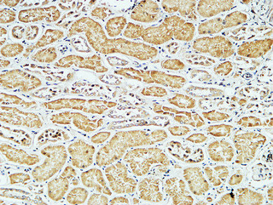
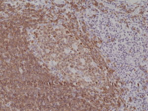
. CD79A was detected in paraffin-embedded section of human tonsil cancer tissue. Heat mediated antigen retrieval was performed in EDTA buffer (pH8.0, epitope retrieval solution). The tissue section was blocked with 10% goat serum. The tissue section was then incubated with 1microg/ml mouse anti-CD79A Antibody (M01047-3) overnight at 4°C. Biotinylated goat anti-mouse IgG was used as secondary antibody and incubated for 30 minutes at 37°C. The tissue section was developed using Strepavidin-Biotin-Complex (SABC) (Catalog # SA1021) with DAB as the chromogen.")
Figure 1. Western blot analysis of CD79A using anti ZO-1 antibody (M01047-3). Electrophoresis was performed on a 5-20% SDS-PAGE gel at 70V (Stacking gel) / 90V (Resolving gel) for 2-3 hours. The sample well of each lane was loaded with 50ug of sample under reducing conditions. Lane 1: human Raji tissue lysates, After Electrophoresis, proteins were transferred to a Nitrocellulose membrane at 150mA for 50-90 minutes. Blocked the membrane with 5% Non-fat Milk/ TBS for 1.5 hour at RT. The membrane was incubated with mouse anti-CD79A antigen affinity purified polyclonal antibody (Catalog # M01047-3) at 0.5 microg/mL overnight at 4°C, then washed with TBS-0.1%Tween 3 times with 5 minutes each and probed with a goat anti-mouse IgG-HRP secondary antibody at a dilution of 1:10000 for 1.5 hour at RT. The signal is developed using an Enhanced Chemiluminescent detection (ECL) kit (Catalog # EK1001) with Tanon 5200 system. A specific band was detected for CD79A at approximately 44KD. The expected band size for CD79A is at 25KD.
Anti-CD79a Antibody Picoband(r) (monoclonal, 4G4)
M01047-3-FITC
ApplicationsWestern Blot, ImmunoHistoChemistry
Product group Antibodies
ReactivityHuman
TargetCD79A
Overview
- SupplierBoster Bio
- Product NameAnti-CD79a Antibody Picoband(r) (monoclonal, 4G4)
- Delivery Days Customer9
- ApplicationsWestern Blot, ImmunoHistoChemistry
- CertificationResearch Use Only
- ClonalityMonoclonal
- Clone ID4G4
- ConjugateFITC
- Gene ID973
- Target nameCD79A
- Target descriptionCD79a molecule
- Target synonymsIGA, IGAlpha, MB-1, MB1, B-cell antigen receptor complex-associated protein alpha chain, CD79a antigen (immunoglobulin-associated alpha), CD79a molecule, immunoglobulin-associated alpha, MB-1 membrane glycoprotein, ig-alpha, membrane-bound immunoglobulin-associated protein, surface IgM-associated protein
- HostMouse
- IsotypeIgG1
- Protein IDP11912
- Protein NameB-cell antigen receptor complex-associated protein alpha chain
- Scientific DescriptionBoster Bio Anti-CD79a Antibody Picoband® (monoclonal, 4G4) catalog # M01047-3. Tested in IHC, WB applications. This antibody reacts with Human. The brand Picoband indicates this is a premium antibody that guarantees superior quality, high affinity, and strong signals with minimal background in Western blot applications. Only our best-performing antibodies are designated as Picoband, ensuring unmatched performance.
- ReactivityHuman
- Storage Instruction-20°C,2°C to 8°C
- UNSPSC12352203
Related products
Product group Antibodies
CD79a Monoclonal AntibodyBSM-33449M
ApplicationsImmunoFluorescence, ImmunoCytoChemistry, ImmunoHistoChemistry, ImmunoHistoChemistry Frozen, ImmunoHistoChemistry Paraffin
ReactivityHuman
TargetCD79A
- SizePrice
Product group Antibodies
ApplicationsFlow Cytometry
TargetCD79A
- SizePrice
Product group Antibodies
Anti-CD79a/IGA Antibody130-10035
ApplicationsELISA
ReactivityHuman
TargetCD79A
- SizePrice
Product group Antibodies
Anti-CD79A AntibodyA101170
ApplicationsWestern Blot, ELISA
ReactivityHuman
- SizePrice
Product group Antibodies

Anti-CD79a [HM47]Ab01318-1.1
ApplicationsImmunoPrecipitation, Western Blot, ImmunoHistoChemistry
ReactivityBovine, Chicken, Equine, Guinea Pig, Human, Mouse, Opossum, Porcine, Primate, Rabbit, Rat
TargetCD79A
- SizePrice
Product group Antibodies
CD79a antibodyGTX131291
ApplicationsWestern Blot
ReactivityHuman
TargetCD79A
- SizePrice
Product group Antibodies
anti-CD79a (C-term) (human), Rabbit Monoclonal (RM297)REV-31-1182-00
ApplicationsWestern Blot, ImmunoHistoChemistry
ReactivityHuman
TargetCD79A
- SizePrice
Product group Antibodies

ApplicationsELISA
ReactivityHuman
TargetCD79A
- SizePrice