


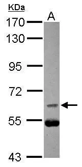
Figure 1. Western blot analysis of Cdc25B using anti-Cdc25B antibody (PA1546). Electrophoresis was performed on a 5-20% SDS-PAGE gel at 70V (Stacking gel) / 90V (Resolving gel) for 2-3 hours. The sample well of each lane was loaded with 30 ug of sample under reducing conditions. Lane 1: Rat Brain Tissue Lysate, Lane 2: Rat Kidney Tissue Lysate, Lane 3: Rat Lung Tissue Lysate. After electrophoresis, proteins were transferred to a nitrocellulose membrane at 150 mA for 50-90 minutes. Blocked the membrane with 5% non-fat milk/TBS for 1.5 hour at RT. The membrane was incubated with rabbit anti-Cdc25B antigen affinity purified polyclonal antibody (Catalog # PA1546) at 0.5 microg/mL overnight at 4°C, then washed with TBS-0.1%Tween 3 times with 5 minutes each and probed with a goat anti-rabbit IgG-HRP secondary antibody at a dilution of 1:5000 for 1.5 hour at RT. The signal is developed using an Enhanced Chemiluminescent detection (ECL) kit (Catalog # EK1002) with Tanon 5200 system. A specific band was detected for Cdc25B at approximately 65 kDa. The expected band size for Cdc25B is at 65 kDa.
. Cdc25B was detected in a paraffin-embedded section of human lung cancer tissue. Heat mediated antigen retrieval was performed in EDTA buffer (pH 8.0, epitope retrieval solution). The tissue section was blocked with 10% goat serum. The tissue section was then incubated with 1 microg/ml rabbit anti-Cdc25B Antibody (PA1546) overnight at 4°C. Biotinylated goat anti-rabbit IgG was used as secondary antibody and incubated for 30 minutes at 37°C. The tissue section was developed using Strepavidin-Biotin-Complex (SABC) (Catalog # SA1022) with DAB as the chromogen.")
Figure 1. Western blot analysis of Cdc25B using anti-Cdc25B antibody (PA1546). Electrophoresis was performed on a 5-20% SDS-PAGE gel at 70V (Stacking gel) / 90V (Resolving gel) for 2-3 hours. The sample well of each lane was loaded with 30 ug of sample under reducing conditions. Lane 1: Rat Brain Tissue Lysate, Lane 2: Rat Kidney Tissue Lysate, Lane 3: Rat Lung Tissue Lysate. After electrophoresis, proteins were transferred to a nitrocellulose membrane at 150 mA for 50-90 minutes. Blocked the membrane with 5% non-fat milk/TBS for 1.5 hour at RT. The membrane was incubated with rabbit anti-Cdc25B antigen affinity purified polyclonal antibody (Catalog # PA1546) at 0.5 microg/mL overnight at 4°C, then washed with TBS-0.1%Tween 3 times with 5 minutes each and probed with a goat anti-rabbit IgG-HRP secondary antibody at a dilution of 1:5000 for 1.5 hour at RT. The signal is developed using an Enhanced Chemiluminescent detection (ECL) kit (Catalog # EK1002) with Tanon 5200 system. A specific band was detected for Cdc25B at approximately 65 kDa. The expected band size for Cdc25B is at 65 kDa.
Anti-Cdc25B Antibody
PA1546
ApplicationsWestern Blot, ImmunoHistoChemistry
Product group Antibodies
ReactivityCanine, Equine, Human, Monkey, Rabbit, Rat
TargetCDC25B
Overview
- SupplierBoster Bio
- Product NameAnti-Cdc25B Antibody
- Delivery Days Customer9
- Application Supplier NoteTested Species: In-house tested species with positive results. Predicted Species: Species predicted to be fit for the product based on sequence similarities. By Heat: Boiling the paraffin sections in 10mM citrate buffer, pH6.0, for 20mins is required for the staining of formalin/paraffin sections. Other applications have not been tested. Optimal dilutions should be determined by end users.
- ApplicationsWestern Blot, ImmunoHistoChemistry
- Applications SupplierIHP, WB, IHC
- CertificationResearch Use Only
- ClonalityPolyclonal
- Concentration500 ug/ml
- Gene ID994
- Target nameCDC25B
- Target descriptioncell division cycle 25B
- Target synonymsMPIP2, M-phase inducer phosphatase 2, CDC25 homolog B, dual specificity phosphatase Cdc25B
- HostRabbit
- IsotypeIgG
- Protein IDP30305
- Protein NameM-phase inducer phosphatase 2
- Scientific DescriptionBoster Bio Anti-M-phase inducer phosphatase 2 Cdc25B Antibody catalog # PA1546. Tested in IHC, WB applications. This antibody reacts with Human, Rat. The brand Picoband indicates this is a premium antibody that guarantees superior quality, high affinity, and strong signals with minimal background in Western blot applications. Only our best-performing antibodies are designated as Picoband, ensuring unmatched performance.
- ReactivityCanine, Equine, Human, Monkey, Rabbit, Rat
- Reactivity SupplierHuman, Rat, Dog, Horse, Monkey, Rabbit
- Storage Instruction-20°C,2°C to 8°C
- UNSPSC12352203
Datasheet
MSDS
Related products

Product group Antibodies
Anti-CDC25B Antibody Picoband(r)A01899-1-CARRIER-FREE
ApplicationsFlow Cytometry, Western Blot, ELISA
ReactivityHuman, Mouse, Rat
TargetCDC25B
- SizePrice
Product group Antibodies
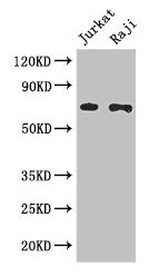
ApplicationsWestern Blot
TargetCDC25B
- SizePrice
Product group Antibodies
CDC25B Polyclonal AntibodyCAC14584
ApplicationsImmunoFluorescence, Western Blot, ELISA, ImmunoHistoChemistry
TargetCDC25B
- SizePrice
Product group Antibodies
CDC25B Recombinant Antibody, Biotin ConjugatedBSM-61332R-BIOTIN
ApplicationsImmunoPrecipitation, Western Blot
TargetCDC25B
- SizePrice
Product group Antibodies
CDC25B AntibodyCSB-PA004995LA01HU
ApplicationsImmunoFluorescence, Western Blot, ELISA, ImmunoHistoChemistry
ReactivityHuman
TargetCDC25B
- SizePrice

Product group Antibodies
Anti-CDC25B AntibodyHPA038892
ApplicationsWestern Blot, ImmunoHistoChemistry
ReactivityHuman
TargetCDC25B
- SizePrice
Product group Antibodies

CDC25B AntibodyLS-C402687
ApplicationsELISA, ImmunoHistoChemistry
ReactivityHuman
TargetCDC25B
- SizePrice
Product group Antibodies
CDC25B antibody [C1C3]GTX113331
ApplicationsWestern Blot
ReactivityHuman
TargetCDC25B
- SizePrice