


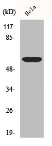
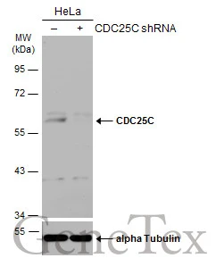
Figure 1. Western blot analysis of Cdc25C using anti-Cdc25C antibody (PB9756). Electrophoresis was performed on a 5-20% SDS-PAGE gel at 70V (Stacking gel) / 90V (Resolving gel) for 2-3 hours. Lane 1: Rat Ovary Tissue Lysate at 50ug, Lane 2: Rat Liver Tissue Lysate at 50ug, Lane 3: HELA Whole Cell Lysate at 40ug, Lane 4: SW620 Whole Cell Lysate at 40ug. After electrophoresis, proteins were transferred to a nitrocellulose membrane at 150 mA for 50-90 minutes. Blocked the membrane with 5% non-fat milk/TBS for 1.5 hour at RT. The membrane was incubated with rabbit anti-Cdc25C antigen affinity purified polyclonal antibody (Catalog # PB9756) at 0.5 microg/mL overnight at 4°C, then washed with TBS-0.1%Tween 3 times with 5 minutes each and probed with a goat anti-rabbit IgG-HRP secondary antibody at a dilution of 1:5000 for 1.5 hour at RT. The signal is developed using an Enhanced Chemiluminescent detection (ECL) kit (Catalog # EK1002) with Tanon 5200 system. A specific band was detected for Cdc25C at approximately 60 kDa. The expected band size for Cdc25C is at 60 kDa.
Figure 1. Western blot analysis of Cdc25C using anti-Cdc25C antibody (PB9756). Electrophoresis was performed on a 5-20% SDS-PAGE gel at 70V (Stacking gel) / 90V (Resolving gel) for 2-3 hours. Lane 1: Rat Ovary Tissue Lysate at 50ug, Lane 2: Rat Liver Tissue Lysate at 50ug, Lane 3: HELA Whole Cell Lysate at 40ug, Lane 4: SW620 Whole Cell Lysate at 40ug. After electrophoresis, proteins were transferred to a nitrocellulose membrane at 150 mA for 50-90 minutes. Blocked the membrane with 5% non-fat milk/TBS for 1.5 hour at RT. The membrane was incubated with rabbit anti-Cdc25C antigen affinity purified polyclonal antibody (Catalog # PB9756) at 0.5 microg/mL overnight at 4°C, then washed with TBS-0.1%Tween 3 times with 5 minutes each and probed with a goat anti-rabbit IgG-HRP secondary antibody at a dilution of 1:5000 for 1.5 hour at RT. The signal is developed using an Enhanced Chemiluminescent detection (ECL) kit (Catalog # EK1002) with Tanon 5200 system. A specific band was detected for Cdc25C at approximately 60 kDa. The expected band size for Cdc25C is at 60 kDa.
Anti-Cdc25C Antibody Picoband(r)
PB9756-CARRIER-FREE
ApplicationsWestern Blot
Product group Antibodies
ReactivityHuman, Rat
TargetCDC25C
Overview
- SupplierBoster Bio
- Product NameAnti-Cdc25C Antibody Picoband(r)
- Delivery Days Customer9
- Application Supplier NoteTested Species: In-house tested species with positive results. Other applications have not been tested. Optimal dilutions should be determined by end users.
- ApplicationsWestern Blot
- CertificationResearch Use Only
- ClonalityPolyclonal
- Concentration500 ug/ml
- Gene ID995
- Target nameCDC25C
- Target descriptioncell division cycle 25C
- Target synonymsCDC25, PPP1R60, M-phase inducer phosphatase 3, CDC25 homolog C, dual specificity phosphatase CDC25C, mitosis inducer CDC25, phosphotyrosine phosphatase, protein phosphatase 1, regulatory subunit 60
- HostRabbit
- IsotypeIgG
- Protein IDP30307
- Protein NameM-phase inducer phosphatase 3
- Scientific DescriptionBoster Bio Anti-Cdc25C Antibody Picoband® catalog # PB9756. Tested in WB applications. This antibody reacts with Human, Rat. The brand Picoband indicates this is a premium antibody that guarantees superior quality, high affinity, and strong signals with minimal background in Western blot applications. Only our best-performing antibodies are designated as Picoband, ensuring unmatched performance.
- ReactivityHuman, Rat
- Storage Instruction-20°C,2°C to 8°C
- UNSPSC12352203
Related products
Product group Antibodies
Anti-CDC25C Antibody144-01672
ApplicationsWestern Blot
ReactivityHuman
TargetCDC25C
- SizePrice
Product group Antibodies
Anti-CDC25C AntibodyA99313
ApplicationsImmunoFluorescence, ELISA, ImmunoHistoChemistry
ReactivityHuman
- SizePrice
Product group Antibodies
Cdc25C Polyclonal AntibodyCAC11791
ApplicationsELISA, ImmunoHistoChemistry
TargetCDC25C
- SizePrice
Product group Antibodies
CDC25C AntibodyCSB-PA001503
ApplicationsWestern Blot, ELISA, ImmunoHistoChemistry
ReactivityHuman
TargetCDC25C
- SizePrice
Product group Antibodies
CDC25C antibodyGTX102809
ApplicationsImmunoFluorescence, Western Blot, ImmunoCytoChemistry, ImmunoHistoChemistry, ImmunoHistoChemistry Paraffin
ReactivityHuman
TargetCDC25C
- SizePrice

Product group Antibodies
Anti-CDC25C AntibodyHPA066991
ApplicationsImmunoCytoChemistry
ReactivityHuman
TargetCDC25C
- SizePrice
Product group Antibodies

CDC25C AntibodyLS-C746941
ApplicationsImmunoFluorescence, Western Blot
ReactivityHuman, Mouse
TargetCDC25C
- SizePrice
Product group Antibodies
References
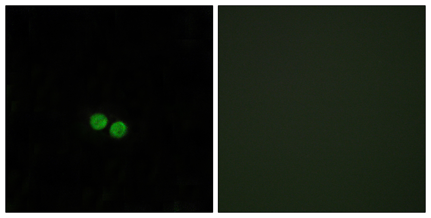
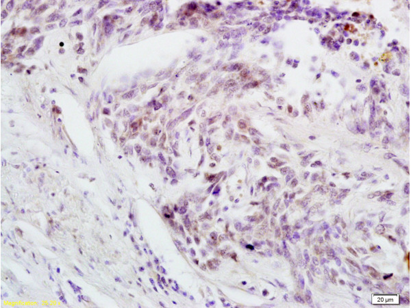
ApplicationsImmunoFluorescence, Western Blot, ELISA, ImmunoCytoChemistry, ImmunoHistoChemistry, ImmunoHistoChemistry Frozen, ImmunoHistoChemistry Paraffin
ReactivityHuman, Mouse, Rat
TargetCDC25C
- SizePrice
Product group Antibodies
Anti-E2F1 AntibodyCAB16720
ApplicationsWestern Blot, ELISA
ReactivityHuman
- SizePrice