

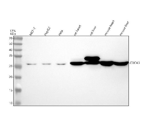
Figure 1. Western blot analysis of CDCA3 using anti-CDCA3 antibody (M10074). Electrophoresis was performed on a 5-20% SDS-PAGE gel at 70V (Stacking gel) / 90V (Resolving gel) for 2-3 hours. The sample well of each lane was loaded with 30 ug of sample under reducing conditions. Lane 1: human MCF-7 whole cell lysates, Lane 2: human HepG2 whole cell lysates, Lane 3: human Hela whole cell lysates, Lane 4: rat heart tissue lysates, Lane 5: rat liver tissue lysates, Lane 6: mouse heart tissue lysates, Lane 7: mouse liver tissue lysates. After electrophoresis, proteins were transferred to a nitrocellulose membrane at 150 mA for 50-90 minutes. Blocked the membrane with 5% non-fat milk/TBS for 1.5 hour at RT. The membrane was incubated with rabbit anti-CDCA3 antigen affinity purified monoclonal antibody (Catalog # M10074) at 0.5 microg/mL overnight at 4°C, then washed with TBS-0.1%Tween 3 times with 5 minutes each and probed with a goat anti-rabbit IgG-HRP secondary antibody at a dilution of 1:5000 for 1.5 hour at RT. The signal is developed using an Enhanced Chemiluminescent detection (ECL) kit (Catalog # EK1002) with Tanon 5200 system. A specific band was detected for CDCA3 at approximately 29 kDa. The expected band size for CDCA3 is at 29 kDa.
Figure 1. Western blot analysis of CDCA3 using anti-CDCA3 antibody (M10074). Electrophoresis was performed on a 5-20% SDS-PAGE gel at 70V (Stacking gel) / 90V (Resolving gel) for 2-3 hours. The sample well of each lane was loaded with 30 ug of sample under reducing conditions. Lane 1: human MCF-7 whole cell lysates, Lane 2: human HepG2 whole cell lysates, Lane 3: human Hela whole cell lysates, Lane 4: rat heart tissue lysates, Lane 5: rat liver tissue lysates, Lane 6: mouse heart tissue lysates, Lane 7: mouse liver tissue lysates. After electrophoresis, proteins were transferred to a nitrocellulose membrane at 150 mA for 50-90 minutes. Blocked the membrane with 5% non-fat milk/TBS for 1.5 hour at RT. The membrane was incubated with rabbit anti-CDCA3 antigen affinity purified monoclonal antibody (Catalog # M10074) at 0.5 microg/mL overnight at 4°C, then washed with TBS-0.1%Tween 3 times with 5 minutes each and probed with a goat anti-rabbit IgG-HRP secondary antibody at a dilution of 1:5000 for 1.5 hour at RT. The signal is developed using an Enhanced Chemiluminescent detection (ECL) kit (Catalog # EK1002) with Tanon 5200 system. A specific band was detected for CDCA3 at approximately 29 kDa. The expected band size for CDCA3 is at 29 kDa.
Anti-CDCA3 Rabbit Monoclonal Antibody
M10074
ApplicationsImmunoFluorescence, Western Blot, ImmunoCytoChemistry
Product group Antibodies
ReactivityHuman, Mouse, Rat
TargetCDCA3
Overview
- SupplierBoster Bio
- Product NameAnti-CDCA3 Rabbit Monoclonal Antibody
- Delivery Days Customer9
- ApplicationsImmunoFluorescence, Western Blot, ImmunoCytoChemistry
- CertificationResearch Use Only
- ClonalityMonoclonal
- Clone IDIAF-3
- Gene ID83461
- Target nameCDCA3
- Target descriptioncell division cycle associated 3
- Target synonymsGRCC8, TOME-1, TOME1, cell division cycle-associated protein 3, gene-rich cluster protein C8, trigger of mitotic entry protein 1
- HostRabbit
- IsotypeIgG
- Protein IDQ99618
- Protein NameCell division cycle-associated protein 3
- Scientific DescriptionBoster Bio Anti-CDCA3 Rabbit Monoclonal Antibody catalog # M10074. Tested in WB, ICC/IF applications. This antibody reacts with Human, Mouse, Rat.
- ReactivityHuman, Mouse, Rat
- Storage Instruction-20°C
- UNSPSC12352203
Datasheet
MSDS
Related products
Product group Antibodies
CDCA3 AntibodyCSB-PA001515
ApplicationsImmunoFluorescence, Western Blot, ELISA, ImmunoHistoChemistry
ReactivityHuman, Mouse, Rat
TargetCDCA3
- SizePrice
Product group Antibodies
Anti-CDCA3 Antibody144-64152
ApplicationsWestern Blot
ReactivityHuman, Mouse
TargetCDCA3
- SizePrice
Product group Antibodies
Anti-CDCA3 AntibodyA89098
ApplicationsWestern Blot
ReactivityHuman, Mouse
- SizePrice

Product group Antibodies
Anti-CDCA3 AntibodyHPA026587
ApplicationsWestern Blot
ReactivityHuman
TargetCDCA3
- SizePrice
Product group Antibodies
CDCA3 Recombinant Antibody, Biotin ConjugatedBSM-61345R-BIOTIN
ApplicationsWestern Blot
ReactivityHuman, Mouse, Rat
TargetCDCA3
- SizePrice
Product group Antibodies

CDCA3 Antibody (aa200-230)LS-C288287
ApplicationsImmunoPrecipitation
ReactivityHuman
TargetCDCA3
- SizePrice