


Immunohistochemical staining of human kidney shows strong membranous positivity in cells in tubules.

Immunohistochemical staining of human kidney shows strong membranous positivity in cells in tubules.
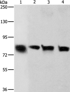
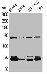
Anti-CDH6 Antibody
HPA007456
ApplicationsWestern Blot, ImmunoHistoChemistry
Product group Antibodies
ReactivityHuman
TargetCDH6
Overview
- SupplierAtlas Antibodies
- Product NameAnti-CDH6 Antibody
- Delivery Days Customer4
- ApplicationsWestern Blot, ImmunoHistoChemistry
- CertificationResearch Use Only
- ClonalityPolyclonal
- ConjugateUnconjugated
- Gene ID1004
- Target nameCDH6
- Target descriptioncadherin 6
- Target synonymsCAD6, KCAD, cadherin-6, cadherin 6, type 2, K-cadherin (fetal kidney)
- HostRabbit
- IsotypeIgG
- Protein IDP55285
- Protein NameCadherin-6
- Scientific DescriptionRecombinant Protein Epitope Signature Tag (PrEST) antigen sequence
- ReactivityHuman
- Storage Instruction-20°C,2°C to 8°C
- UNSPSC41116161
Datasheet
MSDS
Related products
Product group Antibodies
Anti-CDH6 AntibodyA38707
ApplicationsWestern Blot, ImmunoHistoChemistry
ReactivityHuman
- SizePrice

Product group Antibodies
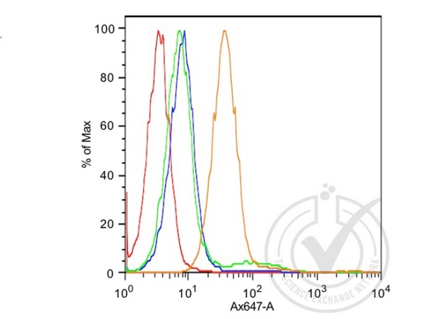
Anti-K Cadherin/CDH6 Antibody Picoband(r)A06353-1-CARRIER-FREE
ApplicationsFlow Cytometry, Western Blot, ELISA, ImmunoHistoChemistry
ReactivityHuman, Mouse, Rat
TargetCDH6
- SizePrice
Product group Antibodies
Anti-CDH6 Antibody144-08109
ApplicationsWestern Blot, ImmunoHistoChemistry
ReactivityHuman, Mouse, Rat
TargetCDH6
- SizePrice
Product group Antibodies

K Cadherin AntibodyABX027844
ApplicationsFlow Cytometry, Western Blot, ELISA, ImmunoHistoChemistry
- SizePrice
Product group Antibodies
K Cadherin Polyclonal AntibodyBS-5823R
ApplicationsFlow Cytometry, Western Blot, ELISA
ReactivityBovine, Equine, Human, Mouse, Rat
TargetCDH6
- SizePrice
Product group Antibodies
CDH6 AntibodyCSB-PA004767
ApplicationsWestern Blot, ELISA
ReactivityHuman, Mouse, Rat
TargetCDH6
- SizePrice
Product group Antibodies

Mouse anti K-Cadherin/Cadherin-6MUB0305P
ApplicationsWestern Blot, ImmunoCytoChemistry, ImmunoHistoChemistry, ImmunoHistoChemistry Frozen, ImmunoHistoChemistry Paraffin
ReactivityHuman, Rat
TargetCDH6
- SizePrice
Product group Antibodies

CDH6 / K Cadherin AntibodyLS-C401314
ApplicationsWestern Blot, ELISA, ImmunoHistoChemistry
ReactivityHuman, Mouse, Rat
TargetCDH6
- SizePrice