


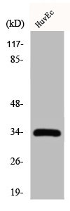
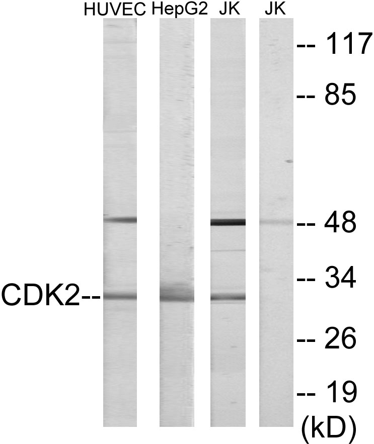
Figure 1. Western blot analysis of Cdk2 using anti-Cdk2 antibody (PB9534). Electrophoresis was performed on a 5-20% SDS-PAGE gel at 70V (Stacking gel) / 90V (Resolving gel) for 2-3 hours. The sample well of each lane was loaded with 30 ug of sample under reducing conditions. Lane 1: human Jurlat whole cell lysates, Lane 2: human HepG2 whole cell lysates, Lane 3: human U2OS whole cell lysates, Lane 4: human K562 whole cell lysates, Lane 5: human T47D whole cell lysates, Lane 6: human CACO-2 whole cell lysates, Lane 7: human 293T whole cell lysates, Lane 8: human PC-3 whole cell lysates. After electrophoresis, proteins were transferred to a nitrocellulose membrane at 150 mA for 50-90 minutes. Blocked the membrane with 5% non-fat milk/TBS for 1.5 hour at RT. The membrane was incubated with rabbit anti-Cdk2 antigen affinity purified polyclonal antibody (Catalog # PB9534) at 0.5 microg/mL overnight at 4°C, then washed with TBS-0.1%Tween 3 times with 5 minutes each and probed with a goat anti-rabbit IgG-HRP secondary antibody at a dilution of 1:5000 for 1.5 hour at RT. The signal is developed using an Enhanced Chemiluminescent detection (ECL) kit (Catalog # EK1002) with Tanon 5200 system. A specific band was detected for Cdk2 at approximately 30 kDa. The expected band size for Cdk2 is at 30, 33 kDa.
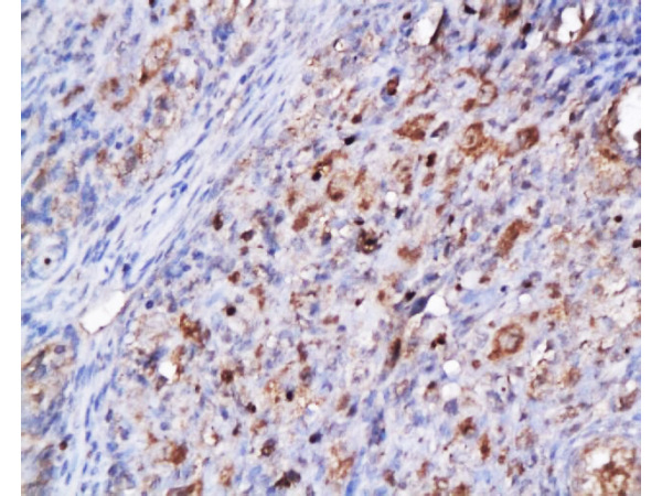
. Cdk2 was detected in a paraffin-embedded section of human invasive urothelial carcinoma of the bladder with squamous differentiation tissue. Heat mediated antigen retrieval was performed in EDTA buffer (pH 8.0, epitope retrieval solution). The tissue section was blocked with 10% goat serum. The tissue section was then incubated with 2 microg/ml rabbit anti-Cdk2 Antibody (PB9534) overnight at 4°C. Peroxidase Conjugated Goat Anti-rabbit IgG was used as secondary antibody and incubated for 30 minutes at 37°C. The tissue section was developed using HRP Conjugated Rabbit IgG Super Vision Assay Kit (Catalog # SV0002) with DAB as the chromogen.")
 and anti-Beta Tubulin antibody (M01857-3). Cdk2 was detected in immunocytochemical section of A549 cell. Enzyme antigen retrieval was performed using IHC enzyme antigen retrieval reagent (AR0022) for 15 mins. The cells were blocked with 10% goat serum. And then incubated with 2 microg/mL rabbit anti-Cdk2 Antibody (PB9534) and mouse anti-Beta Tubulin antibody (M01857-3) overnight at 4°C. DyLight®488 Conjugated Goat Anti-Rabbit IgG (BA1127) and DyLight®594 Conjugated Goat Anti-Mouse IgG (BA1141) were used as secondary antibody at 1:500 dilution and incubated for 30 minutes at 37°C. Visualize using a fluorescence microscope and filter sets appropriate for the label used.")
Figure 1. Western blot analysis of Cdk2 using anti-Cdk2 antibody (PB9534). Electrophoresis was performed on a 5-20% SDS-PAGE gel at 70V (Stacking gel) / 90V (Resolving gel) for 2-3 hours. The sample well of each lane was loaded with 30 ug of sample under reducing conditions. Lane 1: human Jurlat whole cell lysates, Lane 2: human HepG2 whole cell lysates, Lane 3: human U2OS whole cell lysates, Lane 4: human K562 whole cell lysates, Lane 5: human T47D whole cell lysates, Lane 6: human CACO-2 whole cell lysates, Lane 7: human 293T whole cell lysates, Lane 8: human PC-3 whole cell lysates. After electrophoresis, proteins were transferred to a nitrocellulose membrane at 150 mA for 50-90 minutes. Blocked the membrane with 5% non-fat milk/TBS for 1.5 hour at RT. The membrane was incubated with rabbit anti-Cdk2 antigen affinity purified polyclonal antibody (Catalog # PB9534) at 0.5 microg/mL overnight at 4°C, then washed with TBS-0.1%Tween 3 times with 5 minutes each and probed with a goat anti-rabbit IgG-HRP secondary antibody at a dilution of 1:5000 for 1.5 hour at RT. The signal is developed using an Enhanced Chemiluminescent detection (ECL) kit (Catalog # EK1002) with Tanon 5200 system. A specific band was detected for Cdk2 at approximately 30 kDa. The expected band size for Cdk2 is at 30, 33 kDa.
Anti-Cdk2 Antibody Picoband(r)
PB9534-CARRIER-FREE
ApplicationsImmunoFluorescence, Western Blot, ImmunoCytoChemistry, ImmunoHistoChemistry
Product group Antibodies
ReactivityHuman
TargetCDK2
Overview
- SupplierBoster Bio
- Product NameAnti-Cdk2 Antibody Picoband(r)
- Delivery Days Customer9
- Application Supplier NoteTested Species: In-house tested species with positive results. By Heat: Boiling the paraffin sections in 10mM citrate buffer, pH6.0, for 20mins is required for the staining of formalin/paraffin sections. Other applications have not been tested. Optimal dilutions should be determined by end users.
- ApplicationsImmunoFluorescence, Western Blot, ImmunoCytoChemistry, ImmunoHistoChemistry
- CertificationResearch Use Only
- ClonalityPolyclonal
- Concentration500 ug/ml
- Gene ID1017
- Target nameCDK2
- Target descriptioncyclin dependent kinase 2
- Target synonymsCDKN2, p33(CDK2), cyclin-dependent kinase 2, cdc2-related protein kinase, cell division protein kinase 2, p33 protein kinase
- HostRabbit
- IsotypeIgG
- Protein IDP24941
- Protein NameCyclin-dependent kinase 2
- Scientific DescriptionBoster Bio Anti-Cdk2 Antibody Picoband® catalog # PB9534. Tested in IF, IHC, ICC, WB applications. This antibody reacts with Human. The brand Picoband indicates this is a premium antibody that guarantees superior quality, high affinity, and strong signals with minimal background in Western blot applications. Only our best-performing antibodies are designated as Picoband, ensuring unmatched performance.
- ReactivityHuman
- Storage Instruction-20°C,2°C to 8°C
- UNSPSC12352203
Related products
Product group Antibodies
CDK2 AntibodyCSB-PA001533
ApplicationsImmunoFluorescence, Western Blot, ELISA, ImmunoHistoChemistry
ReactivityHuman, Mouse, Rat
TargetCDK2
- SizePrice
Product group Antibodies
Anti-CDK2 AntibodyA96410
ApplicationsImmunoFluorescence, Western Blot, ELISA
ReactivityHuman, Mouse, Rat
- SizePrice

Product group Antibodies
Anti-CDK2 AntibodyAMAB91497
ApplicationsWestern Blot, ImmunoCytoChemistry, ImmunoHistoChemistry
ReactivityHuman
TargetCDK2
- SizePrice
Product group Antibodies

CDK2 Antibody (Preservative Free)LS-C343132
ApplicationsWestern Blot, ELISA
ReactivityHuman
TargetCDK2
- SizePrice
Product group Antibodies
Cdk2 Polyclonal AntibodyCAC07055
ApplicationsImmunoFluorescence, ImmunoPrecipitation, Western Blot, ELISA, ImmunoHistoChemistry
TargetCDK2
- SizePrice
Product group Antibodies
References
CDK2 Polyclonal AntibodyBS-0757R
ApplicationsImmunoFluorescence, Western Blot, ELISA, ImmunoCytoChemistry, ImmunoHistoChemistry, ImmunoHistoChemistry Frozen, ImmunoHistoChemistry Paraffin
ReactivityBovine, Equine, Human, Mouse, Porcine, Rabbit, Rat
TargetCDK2
- SizePrice
Product group Antibodies
Anti-CDK2Y058204
ApplicationsWestern Blot, ELISA, ImmunoHistoChemistry
ReactivityHuman, Mouse, Rat
- SizePrice
Product group Antibodies
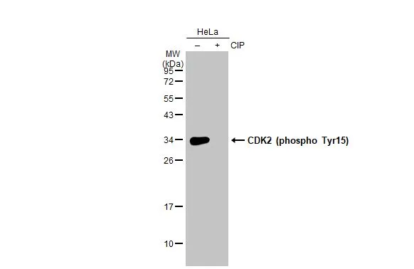
CDK2 (phospho Tyr15) antibodyGTX132802
ApplicationsWestern Blot
ReactivityHuman
TargetCDK2
- SizePrice