


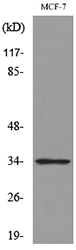
Figure 1. Western blot analysis of CDK4 using anti-CDK4 antibody (PA1428-1, Left) and anti-CDK4 antibody (PB9535, Right). Electrophoresis was performed on a 5-20% SDS-PAGE gel at 70V (Stacking gel) / 90V (Resolving gel) for 2-3 hours. The sample well of each lane was loaded with 30 ug of sample under reducing conditions. Lane 1: human Hela whole cell lysates, Lane 2: human K562 whole cell lysates, Lane 3: rat PC-12 whole cell lysates, Lane 4: mosue NIH/3T3 whole cell lysates, Lane 5: mosue RAW264.7 whole cell lysates. After electrophoresis, proteins were transferred to a nitrocellulose membrane at 150 mA for 50-90 minutes. Blocked the membrane with 5% non-fat milk/TBS for 1.5 hour at RT. The membrane was incubated with rabbit anti-CDK4 antigen affinity purified polyclonal antibody (Catalog # PA1428-1) and rabbit anti-CDK4 antigen affinity purified polyclonal antibody (Catalog # PB9535)at 0.5 microg/mL overnight at 4°C, then washed with TBS-0.1%Tween 3 times with 5 minutes each and probed with a goat anti-rabbit IgG-HRP secondary antibody at a dilution of 1:5000 for 1.5 hour at RT. The signal is developed using an Enhanced Chemiluminescent detection (ECL) kit (Catalog # EK1002)?with Tanon 5200 system. A specific band was detected for CDK4 at approximately 34 kDa. The expected band size for CDK4 is at 34 kDa.
. Cdk4 was detected in a paraffin-embedded section of mouse intestine tissue. Heat mediated antigen retrieval was performed in EDTA buffer (pH 8.0, epitope retrieval solution). The tissue section was blocked with 10% goat serum. The tissue section was then incubated with 1 microg/ml rabbit anti-Cdk4 Antibody (PB9535) overnight at 4°C. Biotinylated goat anti-rabbit IgG was used as secondary antibody and incubated for 30 minutes at 37°C. The tissue section was developed using Strepavidin-Biotin-Complex (SABC) (Catalog # SA1022) with DAB as the chromogen.")
. Cdk4 was detected in a paraffin-embedded section of rat intestine tissue. Heat mediated antigen retrieval was performed in EDTA buffer (pH 8.0, epitope retrieval solution). The tissue section was blocked with 10% goat serum. The tissue section was then incubated with 1 microg/ml rabbit anti-Cdk4 Antibody (PB9535) overnight at 4°C. Biotinylated goat anti-rabbit IgG was used as secondary antibody and incubated for 30 minutes at 37°C. The tissue section was developed using Strepavidin-Biotin-Complex (SABC) (Catalog # SA1022) with DAB as the chromogen.")
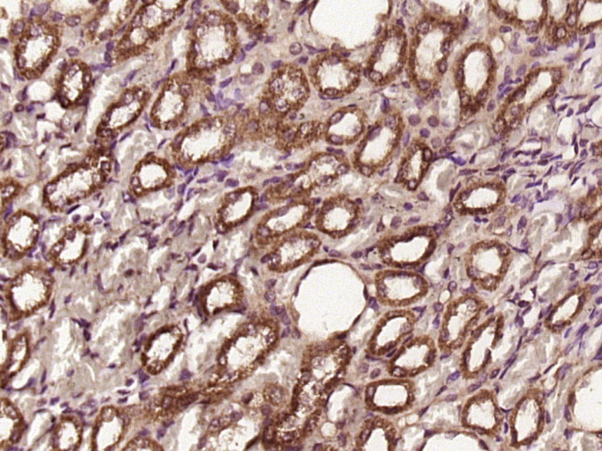
. Cdk4 was detected in a paraffin-embedded section of human lung cancer tissue. Heat mediated antigen retrieval was performed in EDTA buffer (pH 8.0, epitope retrieval solution). The tissue section was blocked with 10% goat serum. The tissue section was then incubated with 1 microg/ml rabbit anti-Cdk4 Antibody (PB9535) overnight at 4°C. Biotinylated goat anti-rabbit IgG was used as secondary antibody and incubated for 30 minutes at 37°C. The tissue section was developed using Strepavidin-Biotin-Complex (SABC) (Catalog # SA1022) with DAB as the chromogen.")
. CDK4 was detected in a paraffin-embedded section of human colon cancer tissue. Heat mediated antigen retrieval was performed in EDTA buffer (pH 8.0, epitope retrieval solution). The tissue section was blocked with 10% goat serum. The tissue section was then incubated with 5 microg/mL rabbit anti-CDK4 Antibody (PB9535) overnight at 4°C. Cy3 Conjugated Goat Anti-Rabbit IgG (BA1032) was used as secondary antibody at 1:500 dilution and incubated for 30 minutes at 37°C. The section was counterstained with DAPI. Visualize using a fluorescence microscope and filter sets appropriate for the label used.")
Figure 1. Western blot analysis of CDK4 using anti-CDK4 antibody (PA1428-1, Left) and anti-CDK4 antibody (PB9535, Right). Electrophoresis was performed on a 5-20% SDS-PAGE gel at 70V (Stacking gel) / 90V (Resolving gel) for 2-3 hours. The sample well of each lane was loaded with 30 ug of sample under reducing conditions. Lane 1: human Hela whole cell lysates, Lane 2: human K562 whole cell lysates, Lane 3: rat PC-12 whole cell lysates, Lane 4: mosue NIH/3T3 whole cell lysates, Lane 5: mosue RAW264.7 whole cell lysates. After electrophoresis, proteins were transferred to a nitrocellulose membrane at 150 mA for 50-90 minutes. Blocked the membrane with 5% non-fat milk/TBS for 1.5 hour at RT. The membrane was incubated with rabbit anti-CDK4 antigen affinity purified polyclonal antibody (Catalog # PA1428-1) and rabbit anti-CDK4 antigen affinity purified polyclonal antibody (Catalog # PB9535)at 0.5 microg/mL overnight at 4°C, then washed with TBS-0.1%Tween 3 times with 5 minutes each and probed with a goat anti-rabbit IgG-HRP secondary antibody at a dilution of 1:5000 for 1.5 hour at RT. The signal is developed using an Enhanced Chemiluminescent detection (ECL) kit (Catalog # EK1002)?with Tanon 5200 system. A specific band was detected for CDK4 at approximately 34 kDa. The expected band size for CDK4 is at 34 kDa.
Anti-Cdk4 Picoband Antibody
PB9535
ApplicationsImmunoFluorescence, Western Blot, ImmunoHistoChemistry
Product group Antibodies
ReactivityHuman, Mouse, Rat
TargetCDK4
Overview
- SupplierBoster Bio
- Product NameAnti-Cdk4 Picoband Antibody
- Delivery Days Customer9
- Application Supplier NoteTested Species: In-house tested species with positive results. By Heat: Boiling the paraffin sections in 10mM citrate buffer, pH6.0, for 20mins is required for the staining of formalin/paraffin sections. Other applications have not been tested. Optimal dilutions should be determined by end users.
- ApplicationsImmunoFluorescence, Western Blot, ImmunoHistoChemistry
- CertificationResearch Use Only
- ClonalityPolyclonal
- Concentration500 ug/ml
- Gene ID1019
- Target nameCDK4
- Target descriptioncyclin dependent kinase 4
- Target synonymsCMM3, PSK-J3, cyclin-dependent kinase 4, cell division protein kinase 4
- HostRabbit
- IsotypeIgG
- Protein IDP11802
- Protein NameCyclin-dependent kinase 4
- Scientific DescriptionBoster Bio Anti-Cdk4 Antibody Picoband® catalog # PB9535. Tested in IF, IHC, WB applications. This antibody reacts with Human, Mouse, Rat. The brand Picoband indicates this is a premium antibody that guarantees superior quality, high affinity, and strong signals with minimal background in Western blot applications. Only our best-performing antibodies are designated as Picoband, ensuring unmatched performance.
- ReactivityHuman, Mouse, Rat
- Storage Instruction-20°C,2°C to 8°C
- UNSPSC12352203
References
- Hou M, Li H, He T, et al. Icariside I reduces breast cancer proliferation, apoptosis, invasion, and metastasis probably through inhibiting IL-6/STAT3 signaling pathway. J Pharm Pharmacol. 2024,76(5):499-513. doi: 10.1093/jpp/rgad103Read this paper
- Sun M, Zhou D, Wu J, et al. Sdy-1 Executes Antitumor Activity in HepG2 and HeLa Cancer Cells by Inhibiting the Wnt/β-Catenin Signaling Pathway. Mar Drugs. 2022,20(2). doi: 10.3390/md20020125Read this paper
- Lu Z, Song W, Zhang Y, et al. Combined Anti-Cancer Effects of Platycodin D and Sorafenib on Androgen-Independent and PTEN-Deficient Prostate Cancer. Front Oncol. 2021,11:648985. doi: 10.3389/fonc.2021.648985Read this paper
- Yin F, Zhao L, Zhang L, et al. Chemopreventive Role of Apigenin against the Synergistic Carcinogenesis of Human Papillomavirus and 4-(Methylnitrosamino)-1-(3-pyridyl)-1-butanone. Biomedicines. 2020,8(11). doi: 10.3390/biomedicines8110472Read this paper
- Zhang Q, Yang X, Luo L, et al. Targeted p21 activation by a new double stranded RNA suppresses human prostate cancer cells growth and metastasis. Am J Transl Res. 2020,12(8):4175-4188.Read this paper
- Fan SH, Xiong QF, Wang L, et al. Glucagon-like peptide 1 treatment reverses vascular remodelling by downregulating matrix metalloproteinase 1 expression through inhibition of the ERK1/2/NF-κB signalling pathway. Mol Cell Endocrinol. 2020,518:111005. doi: 10.1016/j.mce.2020.111005Read this paper
- Wu Y, Ma J, Sun Y, et al. Effect and mechanism of PI3K/AKT/mTOR signaling pathway in the apoptosis of GC-1 cells induced by nickel nanoparticles. Chemosphere. 2020,255:126913. doi: 10.1016/j.chemosphere.2020.126913Read this paper
- Zhao WP, Wang HW, Liu J, et al. JNK/STAT signalling pathway is involved in fluoride-induced follicular developmental dysplasia in female mice. Chemosphere. 2018,209:88-95. doi: 10.1016/j.chemosphere.2018.06.086Read this paper
- Zhang H, Yang Z, Wang J, et al. Wall shear stress promotes intimal hyperplasia through the paracrine H(2)O(2)-mediated NOX-AKT-SVV axis. Life Sci. 2018,207:61-71. doi: 10.1016/j.lfs.2018.05.045Read this paper
- Ren J, Liu Y, Li L, et al. OAMDP, a novel podophyllotoxin derivative, induces apoptosis, cell cycle arrest and autophagy in hepatoma HepG2 cells. Cell Biol Int. 2018,42(2):194-204. doi: 10.1002/cbin.10892Read this paper
Datasheet
MSDS
Related products
Product group Antibodies
ApplicationsWestern Blot, ImmunoHistoChemistry
ReactivityRat
TargetCDK4
- SizePrice
Product group Antibodies
CDK4 Polyclonal AntibodyBS-0633R
ApplicationsFlow Cytometry, ImmunoFluorescence, Western Blot, ELISA, ImmunoCytoChemistry, ImmunoHistoChemistry, ImmunoHistoChemistry Frozen, ImmunoHistoChemistry Paraffin
ReactivityBovine, Human, Mouse, Porcine, Rat
TargetCDK4
- SizePrice
Product group Antibodies
Anti-CDK4 Antibody144-00366
ApplicationsImmunoFluorescence, Western Blot, ImmunoHistoChemistry
ReactivityHuman, Mouse, Rat
TargetCDK4
- SizePrice

Product group Antibodies
Anti-CDK4 AntibodyAMAB91499
ApplicationsWestern Blot, ImmunoCytoChemistry, ImmunoHistoChemistry
ReactivityHuman
TargetCDK4
- SizePrice
Product group Antibodies
Anti-CDK4 AntibodyA98225
ApplicationsWestern Blot, ELISA
ReactivityHuman, Mouse, Rat
- SizePrice
Product group Antibodies
References
CDK4 antibodyGTX102993
ApplicationsImmunoFluorescence, Western Blot, ImmunoCytoChemistry, ImmunoHistoChemistry, ImmunoHistoChemistry Paraffin
ReactivityBovine, Human, Mouse, Rat
TargetCDK4
- SizePrice
Product group Antibodies

CDK4 AntibodyLS-C831727
ApplicationsWestern Blot, ELISA, ImmunoHistoChemistry
ReactivityHuman, Mouse, Rat
TargetCDK4
- SizePrice